Tag Archives: Erin Mills Physiotherapy Ltd.

Vaginismus

Vaginismus

Vaginismus and Physiotherapy

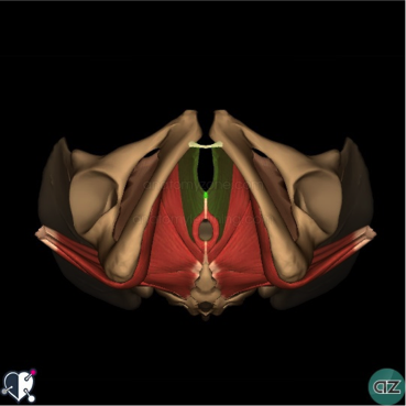
Vaginismus is a painful feeling of discomfort or inability when inserting a tampon, finger, penis or during a doctor’s internal pelvic exam. It occurs when there are involuntary contractions of the muscles in the outer third of the vagina.

Primary Vaginismus: when a woman has never been able to have pain free intercourse due to pelvic floor muscle spasm.

Secondary Vaginismus: pain that develops sometimes later in life after a traumatic event such as childbirth, surgery, or a medical condition.

With Vaginismus, there is usually significant Connective Tissue Dysfunction that needs to be addressed first before any internal work. It is suggested that you follow up the self-help treatment for connective tissue dysfunction before embarking on the stretching exercises with the dilators.

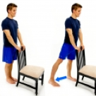
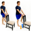
Pelvic floor exercises and Desensitisation techniques

A physiotherapist may be able to teach you pelvic floor exercises, such as squeezing and releasing your pelvic floor muscles, that can help you gain control over the muscles causing the vagina to close involuntarily. Occasionally, a technique called biofeedback may be useful. A small probe is inserted into your vagina. This monitors how well you are doing the exercises by giving you feedback as you do them.
Biofeedback can help with the following:

• Teach you strategies that may help to relax the muscles that are contracting involuntarily
• Teach you appropriate timing of the contraction and relaxation of the pelvic floor muscles.
• Educate you about your condition and why it might be occurring.
• Teach you various strategies for getting the muscles to relax
• help you to Use manual therapy to release the muscles either externally and/or internally that are causing the contractions.

More information about Vaginismus can be found here.

Some general tips about pelvic floor muscle exercises:

• Choose a convenient time and place where you can exercise regularly.
• Put a reminder about pelvic muscle exercises somewhere obvious to you (or set an alarm on your watch or mobile phone).
• Avoid quick pelvic floor muscle exercises where you don’t hold the contraction. This tends to cause an increase in pelvic floor muscle resting tone.
• Be aware that it will take 6 to 8 weeks of regular exercise before you can expect to see improvement. It will take approximately 3 months for the pelvic muscles to strengthen.
• You could try inserting one finger into the vagina to check the strength of your squeeze as you pull in the pelvic muscles. Alternatively, you may be able to feel a pelvic floor contraction if you place the tip of your index finger on the perineal body (on the outside, between the vagina and back passage).

Other tips to reduce Vaginismus:

• If you are overweight, try reducing your weight.
• Try not to become constipated, as a full bowel will put pressure on the bladder. Straining to empty your bowel will weaken the pelvic muscles.
• If your job involves lifting, think of your pelvic muscles as well as your back. Pull up your pelvic muscles when lifting a heavy weight.
• If you smoke, consider quitting. Frequent coughing puts a strain on the pelvic muscles.

If you or someone you know suffers from Vaginismus, please call PhysioNow today! Our specially trained Physiotherapist can start your treatment Now!

Dyspareunia

Dyspareunia and Physiotherapy
Dyspareunia
Dyspareunia is genital pain experienced by women just before, during or after sexual intercourse. Some women have always experienced pain with intercourse from their very first attempt. Other women begin to feel pain with intercourse or cyclically with menstruation. They can also have pain after an injury or infection . Sometimes the pain increases over time. When pain occurs, the woman may be distracted from feeling pleasure and excitement.

Causes

• vaginal dryness from menopause, childbirth, breastfeeding, medications
• skin disorders that cause ulcers, cracks, itching, or burning
• infections, such as yeast or urinary tract infections
• spontaneous tightening of the muscles of the vaginal wall
• endometriosis
• pelvic inflammatory disease
• uterine fibroids
• irritable bowel syndrome
• radiation and chemotherapy

Other factors that affect a woman’s ability to become aroused can also cause dyspareunia.
These factors include:
• stress, which can result in tightened muscles of the pelvic floor
• fear, guilt, or shame related to sex
• self-image or body issues
• medications such as birth control pills
• relationship problems
• cancer, arthritis, diabetes, and thyroid disease
• history of sexual abuse

Symptoms of Dyspareunia

• Pain while inserting a tampon or during penis penetration
• Pain with certain sexual partners
• Deep pain during thrusting
• Burning pain or aching pain
• Throbbing pain, lasting hours after intercourse

Check out this link for more information.

Physiotherapy Treatment for Dyspareunia

Education:

To help describe how the pelvic floor muscles can cause pain. Education can also provide techniques that can be used at home.

Manual Physiotherapy: to mobilise muscle and soft tissue, normalize overactive muscles, improve circulation and desensitize painful areas.

Desensitization therapy: learning vaginal relaxation techniques, such as Kegel exercises, that can decrease pain.

Sex therapy: learning how to re-establish intimacy and improve communication with your partner.

Water-based lubricants rather than petroleum jelly or other oil-based lubricants are preferable. Oil-based lubricants tend to dry the vagina.

Psychologic therapies: Cognitive-behavioral therapy and mindfulness-based cognitive therapy can be helpful.

Pelvic muscle relaxation exercises: used with biofeedback, this can help women with tight pelvic muscles learn to consciously relax tight muscles.

If you or someone you know suffers from dyspareunia, please call today to get started on Physiotherapy treatment! We can help at PhysioNow. Often only a few visits with our specially trained Phyiotherapist can help to get you on the road to recovery.

Hip fracture

Anatomy of Hip
A hip fracture is a break in the upper quarter of the femur (thigh) bone. The extent of the break depends on the forces that are involved. The type of surgery used to treat a hip fracture is based on the bones and soft tissues affected or on the level of the fracture.
Older people are at a higher risk of hip fracture because bones tend to weaken with age (osteoporosis). Multiple medications, poor vision and balance problems also make older people more likely to trip and fall — one of the most common causes of hip fracture.

? Signs and symptoms of a hip fracture include:
• Inability to move immediately after a fall
• Severe pain in your hip or groin
• Inability to put weight on your leg on the side of your injured hip
• Stiffness, bruising and swelling in and around your hip area
• Shorter leg on the side of your injured hip
• Turning outward of your leg on the side of your injured hip

? Causes of Hip fracture
• falling on a hard surface or from a great height.
• blunt trauma to the hip, such as from a car crash.
• diseases such as osteoporosis, which is a condition that causes a loss of bone tissue.
• obesity, which leads to too much pressure on the hip bones.

? Types of Fractures
In general, there are three different types of hip fractures. The type of fracture depends on what area of the upper femur is involved.
Intracapsular Fracture
These fractures occur at the level of the neck and the head of the femur, and are generally within the capsule. The capsule is the soft-tissue envelope that contains the lubricating and nourishing fluid of the hip joint itself.

Intertrochanteric Fracture

This fracture occurs between the neck of the femur and a lower bony prominence called the lesser trochanter. The lesser trochanter is an attachment point for one of the major muscles of the hip. Intertrochanteric fractures generally cross in the area between the lesser trochanter and the greater trochanter. The greater trochanter is the bump you can feel under the skin on the outside of the hip. It acts as another muscle attachment point.

Subtrochanteric Fracture

This fracture occurs below the lesser trochanter, in a region that is between the lesser trochanter and an area approximately 2 1/2 inches below .

In more complicated cases, the amount of breakage of the bone can involve more than one of these zones. This is taken into consideration when surgical repair is considered.

Treatment for hip fracture: usually involves a combination of surgery, rehabilitation and medication.

? Surgery
The type of surgery you have generally depends on the location and severity of the fracture. Are the broken bones properly aligned? (displaced fracture. What is your age? What are your underlying health conditions?

The options include:

• Internal repair using screws. Metal screws are inserted into the bone to hold it together while the fracture heals. Sometimes screws are attached to a metal plate that runs down the upper thigh.

• Partial hip replacement. If the ends of the broken bone are not lined up or damaged, your surgeon may remove the head and neck of the femur and install a metal replacement (prosthesis).

• Total hip replacement. Your upper thigh and your hip socket are replaced with an artificial one (prostheses). Total hip replacement may be a good option if arthritis or a prior injury has damaged your joint. This may have been affecting your hip function even before the fracture.

? Rehabilitation

Rehabilitation is begun as soon as possible after hip fracture surgery, often within a day. The initial goals are to help people retain the level of strength they had before the fracture. You want to keep moving to prevent loss of muscle. You also want to prevent problems that result from bed rest. The ultimate goal is to restore your ability to walk properly without a limp.

Benefits of Rehab

Rehab will help you:

• Restore normal movement in your joint
• Build up strength in the joint and surrounding muscles
• Ease pain and swelling
• Let you get back to your normal activities including walking without a limp
• Help with circulation, particularly right after surgery, so you don’t have problems with blood clots

If you have or someone you know has fractured a hip or had hip replacement surgery, please call PhysioNow. Our experienced physiotherapists would be happy to help with your recovery! Call today to book an appointment!

Trochantric bursitis


Trochanteric bursitis


The Trochanteric Bursa is a fluid filled sac near the hip joint. It cushions between the bones and the muscle and acts as a shock absorber. Trochanteric bursitis is the inflammation of the bursa which is at the outside of the hip.

When this bursa is irritated it causes pain on the outside of the hip and thigh.  Sometimes you may feel the pain down to the knee on the same side.  Another symptom is pain which will stop you from lying on your painful side.  It will be painful when you press on a specific spot on the outside of the hip.  The pain gets worse when climbing stairs, getting up from a low chair getting out of a car.  Sitting with crossed legs hurts.  Both  walking and running may also cause increased pain.

Causes of Trochanteric Bursitis

  • Any direct Injury to the outside of the hip like falling, or bumping into an object.
  • Incorrect posture due to muscle imbalance
  • Leg length difference.
  • Stress of soft tissues surrounding the hip due to hip Osteoarthritis
  • Friction of the ITB (Ilio Tibial Band: a muscle along the side of the hip)
  • Overuse injuries like standing for long periods of time
  • Other diseases which affect the hip like Rheumatoid Arthritis
  • Previous surgeries around the hip
  • Very rarely infection of the bursa

Registered Physiotherapy Treatment

Your Registered Physiotherapist can help you to reduce the pain and inflammation.  They will use electrotherapy modalities, taping, or acupuncture.  They will give you  advice on activity limitations in the early stages.

Later the treatment is focussed towards increasing the flexibility and strength in the muscles.  Your Physiotherapist will  increase the hip joint range of movement and correct your hip joint mechanics.

Trochanteric bursitis

Trochanteric bursitisTrochanteric bursitis

Finally the focus is towards preventing it from happening again.

General Advice on prevention

Your Registered Physiotherapist will look at your posture, hip stability, and core strength.  They will look to see if you  need Orthotics to address any problems with your legs or feet.

 Tips for prevention of Trochanteric Bursitis:

  • Whenever you start a new activity, try to take it slow and build up your activity level gradually.
  • Start with  less force or resistance, less repetitions and progress gradually.
  • Avoid or modify repetitive activities if it is putting any stress on the hip joint
  • Your footwear should be comfortable
  • Maintain flexibility and strength in your lower extremity muscles
  • Improve your core muscle strength

If you or someone you know may have Trochanteric bursitis, call PhysioNow Today!  We would be happy to get you started on your treatment and back to enjoying all the things you love to do!


Vertigo

Benign paroxysmal positional vertigo(BPPV)

Vertigo

Vestibular Systems
• Comprises five sensory organs that provide your brain with information about head position and movements including head rotation, linear movements and static positions of the head relative to gravity
• Five sensory organs including 3 semicircular canals and 2 otoliths


BPPV is a mechanical problem in the inner ear. It occurs when some of the  crystals that are normally embaded in gel in the ear become dislodged.  They then can move into one or more of the 3-fluid filled semicircular canals.

Benign paroxysmal positional vertigo(BPPV) is  one  of the most common causes of vertigo. It creates a false sensation of spinning.
• Benign: it is not life threatening
• Paroxysmal: it comes in sudden, brief spells
• Positional: it gets triggered by certain head positions or movements
• Vertigo: a false sensation of rotational movement


Symptoms
a)      Dizziness
b)      Vertigo (sensation of spinning)
c)       Nausea
d)      Sense of imbalance or unsteadiness
e)      Poor gaze stability
f)       Vision disturbance

Visual Coordination screening


Diagnosis

• The relationship between the inner ears and the eye muscles are what normally allows us to stay focused on our environment while the head is moving.  The dislodged crystals make the brain think you are moving when  you are not. This mistakenly causes the eyes to move, which makes it look like the room is spinning. This is called Nystagmus.

• The Nystagmus will have different characteristics that allow the practitioner to identify which ear the displaced crystals are in and which canal is involved.
• The most common tests are DIX hall pike and Roll test.
• There are two types of  BBPV.  One type, where the loose crystals can move freely in the fluid of the canal(canalithiasis). The more rare type is one where the crystals are thought to be hung up on the bundle of nerves that sense the fluid movement(cupulolithiasis).

Physiotherapy Treatment
• One maneuver that is used for the most common location and type of BPPV is called the Epley maneuver. However , that will not work for all people . Often people have tried the Epley maneuver themselves or had it performed on them without success.
• In the vast majority cases, BPPV can be corrected mechanically by a Registered Physiotherapist. Once your  provider knows which canal is involved and what type it is , we can take you through the appropriate treatment maneuver.

Check out this video for an example of the  Epley Maneuver.  Please do not attempt this yourself until you have been properly assessed and screened by one of our Registered Physiotherapists to see if this maneuver will be appropriate for your condition!

If you have any further questions, please call PhysioNow today! Our experienced Vestibular Physiotherapists would be happy to help you.