Tag Archives: Fracture Treatment

Broken Arm? How to Treat a Distal Humerus Fracture

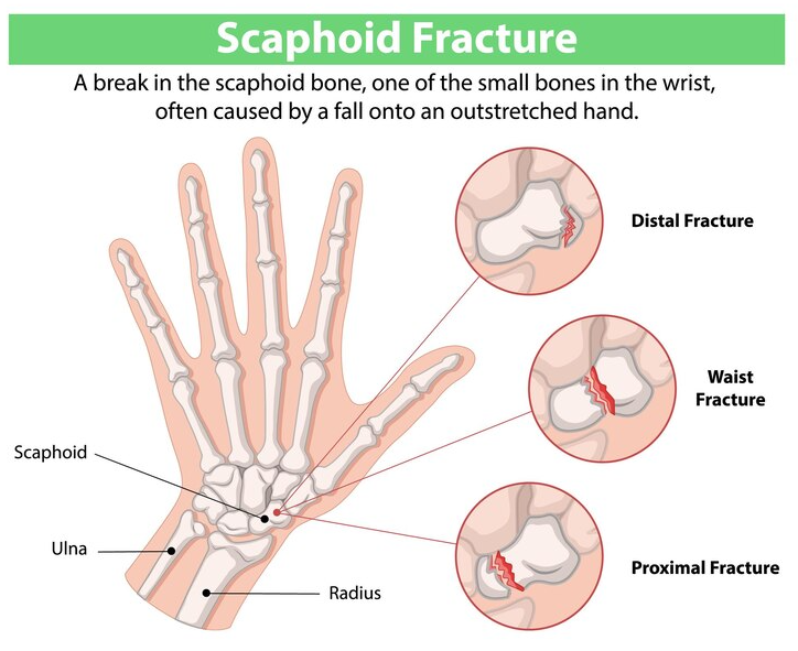
What is a distal humerus fracture?

A distal humerus fracture is a break near the elbow end of the upper arm bone (the humerus). It is one of the more complex types of broken arm injuries because it affects the elbow joint, which is a highly mobile and delicate structure. When this area fractures, it often involves the joint surface, making the injury more complicated than a mid-shaft arm fracture. Fractures can range from simple cracks to severe, multi-fragment breaks, and this will affect healing time and the need for surgical treatment.

Recovery from a humerus fracture requires proper fracture treatment and guided physiotherapy rehabilitation. Otherwise, your arm will not recover to the level it was at prior to injury. For example, untreated injuries frequently result in a loss of range in the elbow, and greatly reduced strength in the affected arm.

What are the common causes of these fractures?

A fall onto an outstretched hand, commonly referred to as a FOOSH injury, is one of the most common ways to fracture the humerus. It occurs a lot in sports where falling is common, such as roller blading, skateboarding and more
  • Falling directly on the elbow or onto an outstretched hand
  • Road traffic accidents
  • Sports injuries, especially contact sports
  • Weak or fragile bones due to osteoporosis, especially in older adults

What are the typical symptoms?

  • Severe pain and swelling around the elbow
  • Bruising or visible deformity near the joint
  • Inability to bend or straighten the elbow
  • Numbness or tingling in the fingers, hand and forearm if nerves are affected

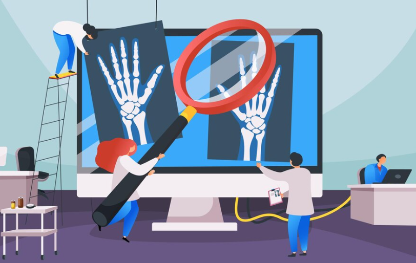
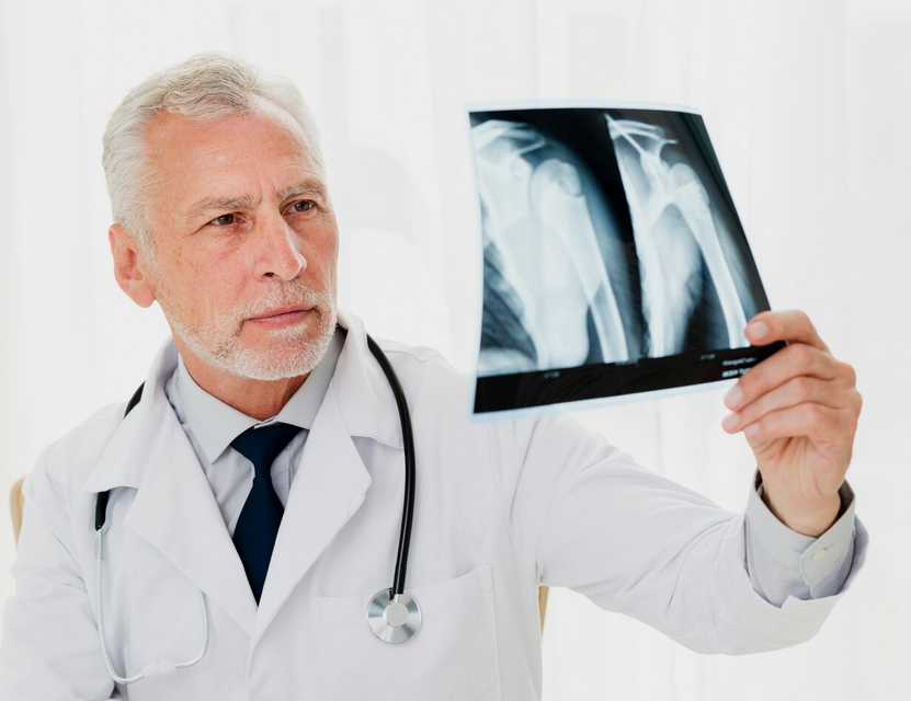
If you experience these symptoms after a fall or impact, you should seek medical attention for an x-ray right away. Early diagnosis and proper protection of a fracture are essential for effective healing.

How is a distal humerus fracture treated?

The best fracture treatment depends on the type and severity of the injury. Treatment usually falls into one of two categories.

Every suspected fracture will be x-rayed upon arrival to the hospital to determine the location and type of fracture. This will help the doctors determine the next steps of your treatment.

Non-surgical (conservative) treatment

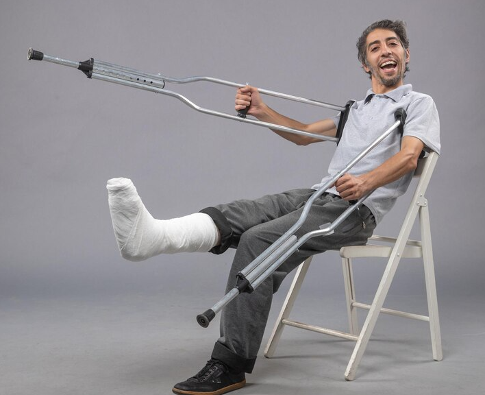
If the fracture is stable or minimally displaced, doctors may recommend immobilization using a cast or brace for several weeks, usually at least 6. This allows the bone to heal naturally while keeping the elbow protected. You will be given some simple range of motion exercises to maintain your shoulder, wrist, and fingers while the elbow heals. After a follow-up x-ray by the fracture clinic, you will be cleared to begin physiotherapy and should start immediately to get the best outcomes.

Surgical treatment

More complex or displaced fractures often require surgery (open reduction and internal fixation). Surgeons use metal plates, screws, or pins to realign and stabilize the bone. Surgery restores joint stability, but physiotherapy afterward is even more important to regain strength and movement.

No matter which approach is taken, starting physiotherapy at the right time is critical to avoid stiffness and recover full function in the arm.

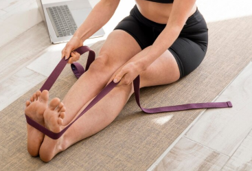
What does physiotherapy look like after a fracture?

Rehabilitation after a distal humerus fracture happens in several key phases. Each phase has specific goals that help your arm recover safely and efficiently.

Phase 1: Immobilization and early healing (0-6 weeks)

Goals: Protect the fracture, reduce pain and swelling, and maintain general fitness as much as possible
Physiotherapy focus (at the fracture clinic):

  • Learn proper sling or brace use
  • Gentle hand, wrist, and shoulder exercises
  • Ice therapy and elevation to control swelling
  • Light isometric (non-moving) muscle contractions to prevent weakness

Phase 2: Early mobilization and strengthening (~6 weeks)

Once your surgeon confirms bone healing through an X-ray, gentle movement can begin
Goals: Regain safe elbow motion and prevent stiffness
Physiotherapy focus (outpatient physiotherapy):

  • Gradual elbow bending and straightening within a pain-free range
  • Gentle forearm rotation exercises (supination and pronation)
  • Continued shoulder and wrist strengthening, gentle forearm, biceps, and triceps strengthening
  • Try to use your hand during normal activities as tolerated, especially if it is your dominant side
  • Manual therapy and soft tissue techniques to reduce stiffness and regain mobility

Phase 3: Strengthening and functional recovery (6–12 weeks)

Goals: Rebuild muscle strength, coordination, and endurance for daily tasks
Physiotherapy focus (outpatient physiotherapy):

  • Active range of motion through full movement
  • Gentle resistance training using light weights or bands
  • Upper limb and shoulder blade strengthening
  • Proprioception (joint awareness) training
  • Gradual return to activities like dressing, typing, and cooking

Phase 4: Advanced strength and return to activity (3–6 months)

Goals: Restore full function, stability, and confidence for work, sports, or hobbies
Physiotherapy focus:

  • Progressive resistance and endurance exercises
  • Sport- or job-specific movement training
  • Weight-bearing and closed-chain exercises for elbow stability
  • Posture and ergonomic guidance for long-term recovery

What challenges can happen during recovery?

Recovering from a distal humerus fracture takes patience and consistent effort. Some common challenges include:

  • Elbow stiffness and a loss of range (usually straightening) caused by scar tissue or long periods of immobilization
  • Weakness from muscle loss during healing
  • Nerve irritation, especially of the ulnar nerve, leading to tingling in the ring and little fingers. Some people recover from this over months to years, some people have permanently altered sensation
  • Fear of movement or anxiety about re-injury leading to lack of use in the arm, stalling recovery

Thankfully, a qualified physiotherapist can help you overcome these issues through a supervised exercise program, manual therapy, and education to rebuild your confidence and function. They will guide you through each step of the process and make a personalized plan that supports your unique goals and situation. If you’re recovering from elbow pain, or a recent fracture, look for PhysioNow! We have many expert Physiotherapists ready to help you recover. We have 10 locations across the west GTA including Burlington, Oakville, Mississauga, and Etobicoke, so please stop in! Alternatively, give us a call at 289-724-0448, or click here to book your first appointment!

Scaphoid Fractures: A Physiotherapy Guide

What is a scaphoid fracture?

A scaphoid fracture is one of the most common types of wrist fractures, particularly among young adults and athletes. The scaphoid bone is one of the eight small bones in the wrist, located in the middle of the joint closer to the thumb side. Functionally, it helps to connect the forearm bones (radius and ulna) to the hand bones (metacarpals). A fracture of the scaphoid typically occurs when an individual falls onto an outstretched hand (commonly shortened to FOOSH), causing the wrist to bend backward in an awkward position. The impact can break the bone, leading to pain, swelling, and difficulty moving the wrist. Unfortunately, due to its location and blood supply, scaphoid fractures can be tricky to diagnose and treat, and failure to do so properly may lead to long-term complications.

How do scaphoid fractures happen?

Activities that have a lot of high impact and weightbearing on the wrist are more susceptible to a fracture

The primary cause of a scaphoid fracture is trauma to the wrist, often from falling onto an outstretched hand. Some common scenarios include:

  • Sports injuries: Athletes in contact sports such as football, basketball, or acrobatic sports like gymnastics are particularly prone to wrist injuries
  • Motor vehicle accidents: Sudden impacts can cause severe trauma to the wrist, leading to fractures
  • Repetitive stress: In some cases, repeated stress or pressure on the wrist may contribute to stress fractures of the scaphoid bone

Additionally, certain risk factors can make someone more prone to a scaphoid fracture, including:

  • Age: Young adults, especially those between 18-30 years old, are more likely to experience this type of fracture
  • Bone density: People with low bone density or osteoporosis may be more susceptible
  • Activity level: Individuals involved in high-impact or contact sports have a higher risk of injury

What are the symptoms of a scaphoid fracture?

 A scaphoid fracture is often difficult to diagnose at first because symptoms may resemble those of other wrist injuries. Common symptoms include:

  • Pain in the wrist: The pain is usually felt on the thumb side of the wrist and may be more intense when the wrist is moved or when pressure is applied to the area
  • Swelling and bruising: Swelling is typically visible around the base of the thumb, and bruising may appear later
  • Reduced range of motion: It may be difficult to move the wrist or thumb without sharp pain
  • Tenderness: Pressing on the area near the base of the thumb is very sore

How can this type of fracture be diagnosed?

To diagnose a scaphoid fracture, doctors will typically perform a physical examination and review the patient’s medical history, including any recent falls or trauma. X-rays are the primary imaging tool used to diagnose a scaphoid fracture, but it’s important to note that fractures may not always be visible immediately after the injury. Sometimes, a second set of X-rays taken a week or two after the injury is required to detect any changes. Unfortunately, scaphoid fractures are unique, and some may never be detectable on x-rays, called occult scaphoid fractures. In these cases, the physical exam and symptoms can usually inform the diagnosis of a fracture.

How is a scaphoid fracture treated?

Treatment for a scaphoid fracture depends on the severity of the break and its location. Generally, treatment options include:

  1. Conservative treatment: Non-displaced fractures (where the bone fragments remain in alignment) are often treated with immobilization using a cast or a splint. This method is typically effective for stable fractures, and the cast is worn for about 6-12 weeks. Here, the wrist is kept in a position that limits movement to allow the bone to heal properly
  2. Surgical treatment: Displaced fractures (where the bone fragments are out of alignment) often require surgical intervention. Typically, surgery involves realigning the bones and using screws, pins, or plates to hold the fragments in place during healing. Also, surgery may be necessary if there’s a concern about the blood supply to the scaphoid bone, which can be disrupted by a fracture. This is an important complication to watch for as it can lead to a serious issue if untreated, called avascular necrosis (AVN)
  3. Rehabilitation: After the cast or surgical treatment, physical therapy is recommended to help restore the wrist’s strength and range of motion. For example, rehabilitation exercises can aid in reducing stiffness, improving flexibility and function of your wrist and hand

Initially, range of motion exercises after immobilization should focus on active-assisted ROM as the hand and wrist will be stiff. These exercises should focus on the wrist and thumb; however, the fingers, elbow and shoulder also need to be considered as they may be affected. If the full range is still restricted, manual therapy can be applied in the form of joint mobilizations to the radio-carpal joint, radio-ulnar and carpal joints. Furthermore,, other forms of manual therapy may also be helpful to reduce any residual swelling or pain such as soft tissue work or massage techniques.

What can go wrong with a fracture?

Although most scaphoid fractures heal successfully with proper treatment, complications can arise. For example, some potential complications include:

  • Nonunion: If the fracture does not heal properly, the bone may fail to unite, leading to chronic pain and dysfunction
  • Avascular necrosis (AVN): The scaphoid bone has a relatively poor blood supply, and fractures, especially those in the proximal part of the bone, can disrupt blood flow. Consequently, this can lead to the death of the bone tissue, which may result in permanent wrist damage
  • Osteoarthritis: Chronic wrist pain or deformity following a scaphoid fracture increases the risk of developing arthritis in the joint

How can I minimize the risk of a fracture?

While some scaphoid fractures are due to accidents or unavoidable trauma, there are steps you can take to reduce your risk:

  • Wear protective gear: When engaging in sports or high-risk activities, wearing wrist guards or other protective equipment can reduce the chances of injury
  • Strengthen the wrist: Regularly performing exercises that strengthen the muscles and ligaments around the wrist can provide more stability and help prevent injuries
  • Fall prevention: Practicing good techniques and safety measures to avoid falling, such as using proper footwear can be especially beneficial

Looking for help?

If you are dealing with a wrist pain, post-fracture issues, or any other type if injury, look for PhysioNow! We have many experienced Registered Physiotherapists ready to help you get better! With 10 different clinics across the GTA from Burlington, Oakville, Mississauga and Etobicoke, our offices are in prime locations to serve you. Contact us if you have any questions and book with us today for your first assessment and treatment.

Fibular Fracture: I Broke my Leg!

What is a fibular fracture?

The fibula is one of two bones in the lower leg, the other being the tibia. Fractures of the tibia and fibula are common in those who have low bone density, specific diseases or anatomical features resulting in changes in bone structure. Also,the likelihood may also depend on the activity level, sports and types of training individuals do. 

Fibular fractures can happen anywhere along the shaft of the bone. Fortunately, due to the smaller size and degree of weight bearing of the fibula relative to the tibia, a fibular fracture is considered less severe. 

How do these fractures happen?

Usually, these fractures are caused by traumatic force to the bone.

  • High impact trauma: For example, these can be from a car crash, pedestrians being hit by a vehicle, falls from heights and more
  • Sports injuries: Especially, in contact sports like football or hockey. Alternatively, a sudden twist or rotational force can fracture the fibula even without a direct blow

What does a fibular fracture look like?

A fracture will result in a sharp, severe pain on the leg which worsens when you put weight on it

Symptoms of a fibular fracture may include:

  1. Pain and tenderness along the outside of the leg
  2. Swelling on the outside or sometimes the entire lower leg
  3. Visible deformity
  4. Inability or difficulty to bear weight on affected leg
  5. Numbness and coldness in feet 

When do I start Physiotherapy Treatment?

After being in a cast for several weeks and being cleared by the fracture clinic, you should see a Physiotherapist for Fracture Rehabilitation. Oftentimes, the knee and ankle become stiff, and the muscles weaken from disuse. Consequently, it is important to begin Physiotherapy Treatment as soon as you can. First, you will focus on regaining the range of motion, strength, and coordination in the lower leg. Simultaneously, you will work on restoring your normal walking pattern and functional abilities like weightbearing, balancing, squatting and stairs. Below is an example of a general rehabilitation program with Physiotherapy after a fibular fracture.

Phase 1: Protection (0-6 weeks)

  1. Patient will be in a cast for about 6 weeks and may be restricted in putting weight on the leg
  2. Encourage leg elevation
  3. Focus on ankle pumps, upper extremity and core strengthening

Phase 2: Early mobility and strengthening (6-8 weeks)

Exercises help increase the range of motion by pushing the ankle into dorsiflexion
  1. Focus on restoring range of motion in all the lower extremity joints
  2. Strong emphasis on ankle dorsiflexion range (bringing your ankle up)
  3. Focus on foot intrinsic muscle strengthening
  4. Start strengthening exercises for the musculature around the ankle, knee, and hip
  5. Gradual progression of weight bearing back to normal
  6. Gait retraining

Phase 3: Continue strengthening (8-12+ weeks)

Box jumps are a high impact exercise that challenges the entire lower body. Consequently, they require a strong foundation of strength before beginning.
  1. Advanced strengthening exercises including resistance training ex. weighted squats, lunges
  2. Start incorporating higher impact activities such as jogging
  3. Progress to plyometric (jumping) and exercises in multiple planes

It is important to note that not everybody will move through the phases of rehabilitation at the same pace. Thus, it is important to have supervision from a trained professional like a Physiotherapist. Ultimately, this will ensure that you are progressing appropriately and minimize your risk of reinjury.

Get started with PhysioNow!

If you are dealing with a fracture or any other types of leg pain, look for PhysioNow! We have many experienced clinicians including Physiotherapists, Massage Therapists, Manual Osteopaths, and Athletic Therapists to assist you in your recovery. We have 10 locations with free and accessible parking located across the GTA. Book with PhysioNow today for your first assessment and treatment!

Images by FreePik