Tag Archives: sports taping

Peroneal tendinopathy

Peroneal tendinopathy

Peroneal tendinopathy

Peroneal tendinopathy or peroneal tendonitis is characterized by an aching pain and swelling in the perineal tendons. These are located in the lower, outside portion of the ankle. A tendon is soft-tissue that attaches a muscle to a bone. The muscles involved in this condition are the 2 peroneal muscles in the lower leg, called the peroneus longus and the peroneus brevis.
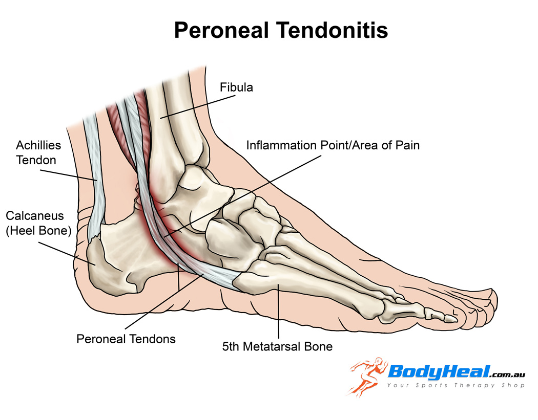
Anatomy

?There are two peroneal tendons that run along the back of the fibula. The first is called the peroneus brevis. The term “brevis” implies short.  It is called this because it has a shorter muscle and starts lower in the leg. It then runs down around the back of the bone called the fibula on the outside of the leg and connects to the side of the foot.  The peroneus longus takes its name because it has a longer course. It starts higher on the leg and runs all the way underneath the foot to connect on the other side of the foot. Both tendons, however, share the major job of turning the ankle to the outside. The tendons are held in a groove behind the back of the fibula bone.

Causes of Peroneal Tendonitis

  • A sudden increase in weight bearing activities, particularly walking, running or jumping
  • Inadequate or unsupportive footwear
  • Muscle imbalances of the lower limb
    Poor lower limb biomechanics
  • Incomplete rehabilitation following an acute ankle injury, such as an ankle sprain

Symptoms of Peroneal Tendinopathy

  • Gradual worsening pain over the outside of the ankle
  • Pain during and/or after weight bearing activities
    Pain with turning the foot in and/or out
  • Instability around the ankle when weight bearing

Diagnosis

A full examination from a physiotherapist can be all thats needed to diagnose peroneal tendonitis
Patients with this condition usually experience pain behind the outside ankle during activities putting stress on the perineal tendons. Pain can also be noticed following these activities or following a rest period. This may be noticed especially upon waking in the morning. There may be swelling when the injury first happens. There will also be pain when testing resisted foot movements.  Stretches into various positions of the foot inversion, and resisted movements can cause pain behind the outside ankle.
Diagnosis may be confirmed with an MRI scan or ultrasound investigation
a diagnostic Ultrasound may be used for detecting all types of peroneal injuries.

What else could it be?:

Symptoms of peroneal tendinopathy mimic various other conditions of the ankle joint. So, before diagnosing peroneal tendinopathy we should rule out other possible injuries by doing the following tests:
Ankle Sprain: ligament testing by the Physiotherapist
Ankle fractures: special tests by the Physiotherapist
Os trigonum syndrome: MRI, physiotherapy testing
Chronical lateral ankle pain with other cause: MRI
Longitudinal peroneal tendon tear: MRI
Peroneal subluxation: ultrasonography, CT, MRI or peroneal tenography
Flexor Hallucis longus tendon injury

Physiotherapy rehabilitation

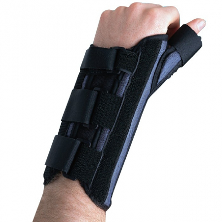
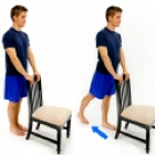
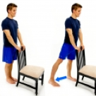
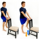
Treatment for peroneal tendonitis includes a program of stretching, strengthening, mobilisation and manipulation. It also includes proprioceptive exercises, icing, ankle bracing or k-taping during contact sports. If symptoms are severe, a cast or ROM boot immobilization may be worn for 10-20 days. After symptoms resolve, you will begin a progressive rehabilitation programme along with a gradual increase to full activity.

The use of a biomechanical ankle platform (BAPS), deep tissue friction massage, ultrasound electric stimulation can also be included in the physiotherapy
Also, shock wave therapy (ESWT), acupuncture is used to treat tendinopathy. But there is only limited evidence from studies for these treatments.
There is evidence for using manual therapy, specifically the lateral calcaneal glide.

If you have any further queries please call PhysioNow. Our experienced physiotherapists would be happy to help you. Call Today to get started 289-724-0448.!

De Quervain’s Disease

De Quervain’s Disease/ Texting thumb.

De Quervain's Disease

De Quervain’s Disease

De Quervain’s Disease or nowadays known as texting thumb is a painful inflammation of tendons in the thumb that extend to the wrist. The rubbing of the inflamed tendon against the canal it passes through causes pain at the base of the thumb and into the lower arm. It is commonly seen in females over 40 years of age.

Causes of De Quervain’s Disease

1. Simple strain injury to the tendon.
2. Repetitive motion injury. Workers who perform rapid repetitive activities involving pinching, grasping, pulling or pushing are at increased risk. Specific activities including intensive mousing, trackball use, and typing. Other activities including bowling, golf, fly-fishing, piano-playing, sewing, and knitting can also cause De Quervain’s Disease.
3. Frequent causes of De Quervain’s Disease include stresses such as lifting young children into car seats, lifting heavy grocery bags by the loops, and lifting gardening pots .
4. De Quervain’s Disease often occurs during and after pregnancy. Factors may include hormonal changes, fluid retention and more lifting.
5. Rheumatoid arthritis.

De Quervain's Disease

De Quervain’s Disease/ Texting Thumb

Onset and Symptoms of De Quervain’s Disease

Onset can be gradual or sudden. Pain is felt along the back of the thumb. There can be Pain directly over the thumb tendons, and pain may travel into the thumb or up the forearm. The bottom of the thumb or the side of the wrist might also be sore or swollen.
It may be hard and painful. Symptoms may get worse when the thumb is moved, particularly when pinching or grasping things. Some people also have swelling and pain on the side of the wrist at the base of the thumb. The back of the thumb and index finger may also feel numb. People might experience a funny sound like a squeak, crackle, snap, or creak when they move the wrist or thumb.
If the condition is not well addressed, the pain can spread up your forearm or down into your thumb.

How is De Quervain’s Disease diagnosed?

De Quervain’s Disease is diagnosed based on history and physical examination. X-rays, or ultrasound may be used to rule out other causes of pain.The Physiotherapist may use special tests to help diagnose De Quervain’s. More information can be found here.

Physiotherapy Treatment

Your Physiotherapist will likely recommend that you wear a specific wrist splint with a thumb spica for 4 to 6 weeks . PhysioNow carries these wrist splints in stock. You’ll also need to stop doing activities that worsen the condition.

Wrist brace with thumb spica

De Quervain’s Disease

The physiotherapist after a thorough assessment could choose different therapy approaches to help with the swelling, pain, and function.
These treatments may include Ultrasound, K-taping, acupuncture, and manual therapy. Other Physiotherapy treatments may include specific exercises focusing on range of motion, strength, and flexibility. These would be given for a safe and effective return back to function.

Recovery times vary depending on your age, general health, and how long you’ve had the symptoms.
If your disease has developed gradually, it’s often tougher to treat. So, it may take you longer to get relief. Your doctor may give you anti-inflammatory medication, or may inject the area with steroids to curb pain and swelling.

In our experience at PhysioNow, more than 99% of people with De Quervain’s Disease get better with Physiotherapy treatment provided. If however, you are one of the outliers, your doctor may recommend surgery. The operation would release the tendon’s tight covering so that the tendon could move smoothly. It’s an outpatient procedure, which means you go home afterward. Your doctor will recommend physiotherapy after surgery which includes an exercise program to strengthen your thumb and wrist.

If you or someone you know suffers from De Quervain’s Disease, please call us today. Our skilled Physiotherapists can Help!

Trochantric bursitis


Trochanteric bursitis


The Trochanteric Bursa is a fluid filled sac near the hip joint. It cushions between the bones and the muscle and acts as a shock absorber. Trochanteric bursitis is the inflammation of the bursa which is at the outside of the hip.

When this bursa is irritated it causes pain on the outside of the hip and thigh.  Sometimes you may feel the pain down to the knee on the same side.  Another symptom is pain which will stop you from lying on your painful side.  It will be painful when you press on a specific spot on the outside of the hip.  The pain gets worse when climbing stairs, getting up from a low chair getting out of a car.  Sitting with crossed legs hurts.  Both  walking and running may also cause increased pain.

Causes of Trochanteric Bursitis

  • Any direct Injury to the outside of the hip like falling, or bumping into an object.
  • Incorrect posture due to muscle imbalance
  • Leg length difference.
  • Stress of soft tissues surrounding the hip due to hip Osteoarthritis
  • Friction of the ITB (Ilio Tibial Band: a muscle along the side of the hip)
  • Overuse injuries like standing for long periods of time
  • Other diseases which affect the hip like Rheumatoid Arthritis
  • Previous surgeries around the hip
  • Very rarely infection of the bursa

Registered Physiotherapy Treatment

Your Registered Physiotherapist can help you to reduce the pain and inflammation.  They will use electrotherapy modalities, taping, or acupuncture.  They will give you  advice on activity limitations in the early stages.

Later the treatment is focussed towards increasing the flexibility and strength in the muscles.  Your Physiotherapist will  increase the hip joint range of movement and correct your hip joint mechanics.

Trochanteric bursitis

Trochanteric bursitisTrochanteric bursitis

Finally the focus is towards preventing it from happening again.

General Advice on prevention

Your Registered Physiotherapist will look at your posture, hip stability, and core strength.  They will look to see if you  need Orthotics to address any problems with your legs or feet.

 Tips for prevention of Trochanteric Bursitis:

  • Whenever you start a new activity, try to take it slow and build up your activity level gradually.
  • Start with  less force or resistance, less repetitions and progress gradually.
  • Avoid or modify repetitive activities if it is putting any stress on the hip joint
  • Your footwear should be comfortable
  • Maintain flexibility and strength in your lower extremity muscles
  • Improve your core muscle strength

If you or someone you know may have Trochanteric bursitis, call PhysioNow Today!  We would be happy to get you started on your treatment and back to enjoying all the things you love to do!


Foot Pain Treatment Mississauga

strong>Foot Pain Treatment Mississauga
Trending Now: Blue Jays’ Jose Batista on 15 day disabled list

Foot Pain Treatment Mississauga

Blue Jays’ Jose Batista hurt his big toe during a June 16 game against Philadelphia. After running into the outfield wall, Bautista had to leave the game early because of pain. An MRI showed that he had suffered a sprain of his left big toe. He had to be in a walking boot for the time being. It may seem like it’s an excessive treatment plan for just a simple sprain. In reality sprains can sometimes be more complicated than a fracture.

What is a Ligament?

A ligament is a soft tissue structure that connects two bones together. Ligaments do not receive a good blood supply like muscles or tendons and as a result, they cannot heal as well.

Types of Sprains

A sprain is defined as a stretch or tear of a ligament.
Furthermore, sprains can be classified into three groups based on the severity of injury. A grade I sprain means that there is mild damage to the ligament and the joint is stable. A grade II sprain means that there is a partial tear in the ligament and the joint is overall less stable or loose. The most severe type of sprain is classified as grade III and it means that the ligament has completely torn or ruptured and the joint has lost stability.

How can a Registered Physiotherapist help with Foot Pain treatment Mississauga?

A Registered Physiotherapist helps with all three types of sprains. The treatment for sprains starts with rest and reducing inflammation. Depending on the type of sprain, it may be necessary for immobilization in order for this to occur. For example a grade II sprain you can get a removable walking boot. A grade III sprain you can get a cast. Once inflammation is reduced and pain is better, treatment seeks to improve range of motion and strength and a return to activity or sport.

Since Jose Batista has been wearing a walking boot, it seems that his injury is at least a grade II. Considering the poor blood supply of ligaments and the severity of his particular sprain it is definitely best for him to be out of the game for at least 2 weeks. Proper foot pain treatment Mississauga in the early phase of injury is very important so that the ligament is well protected and given the best chance for healing. Once the walking boot comes off, his injury will be re-evaluated and the Blue Jays’ medical team will decide on his further treatment plan and eventual return to play.

Jose Bautista’s toe injury improving

If you have been injured, call PhysioNow today! They can help with Foot Pain treatment Mississauga!

Osgood-Schlatter disease

Osgood-Schlatter disease

Osgood-Schlatter disease can cause a painful lump below the kneecap in children and adolescents experiencing growth spurts during puberty.
Osgood-Schlatter disease occurs most often in children who participate in sports that involve running, jumping and swift changes of direction — such as soccer, basketball, figure skating and ballet.
Age ranges differ by sex because girls experience puberty earlier than do boys. Osgood-Schlatter disease typically occurs in boys ages 13 to 14 and girls ages 11 to 12. The condition usually resolves on its own, once the child’s bones stop growing.

Osgood-Schlatter Disease

SYMPTOMS
Most parents call the doctor after their child complains of intermittent pain over several months. The pain may be anywhere from mild and felt only during activity to severe and constant.
Other symptoms may include:
• pain that worsens with exercise
• relief from pain with rest
• swelling or tenderness under the knee and over the shinbone
• limping after exercise
• tightness of the muscles surrounding the knee (the hamstring and quadriceps muscles)

Osgood-Schlatter Disease

Causes
• During activities that involve a lot of running, jumping and bending — such as soccer, basketball, volleyball and ballet —child’s thigh muscles (quadriceps) pull on the tendon that connects the kneecap to the shinbone.
• This repeated stress can cause the tendon to pull away from the shinbone a bit, resulting in the pain and swelling associated with Osgood-Schlatter disease. In some cases, your child’s body may try to close that gap with new bone growth, which can result in a bony lump at that spot.

DIAGNOSIS
• A doctor will perform a physical exam and check your child’s knee for swelling, pain, and redness. This will usually provide the doctor with enough information to make an Osgood-Schlatter disease diagnosis.
• In some cases, the doctor may want to perform a bone X-ray to rule out other potential causes of knee pain.

TREATMENT
Treatment typically involves:
• icing the affected area two to four times a day, or after doing physical activity
• taking over-the-counter pain relievers, such as ibuprofen or acetaminophen
• resting the knee or reducing physical activity
• wrapping the knee or wearing a knee brace
• stretching
physiotherapy

FREQUENTLY ASKED QUESTIONS

1.What about playing sports and other activities?

Sport or physical activity is not likely to cause any permanent damage, but may make the pain worse. However, it is often sporty teenagers who develop Osgood-Schlatter disease. If the pain is affecting your ability to do your sport, difficult decisions may have to be made about training schedules and sporting commitments.

2.Is it possible to prevent Osgood-Schlatter disease?

Yes, it’s possible to prevent Osgood-Schlatter disease by limiting stress activities involving the knees. Such sports would include basketball, volleyball, ballet, gymnastics, and high jump. All of these sports require sudden explosive activation of the quadriceps muscles. A proper stretching routine involving the quadriceps and hamstring muscle groups may be helpful to arrest the onset of Osgood-Schlatter disease.

3.Is this disease permanent?

Osgood-Schlatter disease usually resolves on its own once a growth spurt ends. Until then, treatment is focused on relieving symptoms, such as knee pain and swelling.

• Overall Osgood-Schlatter disease could be a debilitating condition if it remains untreated. PhysioNow Mississauga has excellent Registered Physiotherapists who can help your child and get him or her back to their sports career. Call Today 289-724-0448.