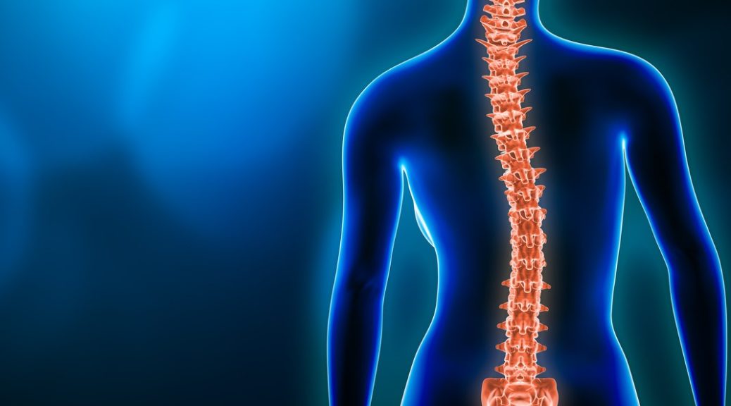
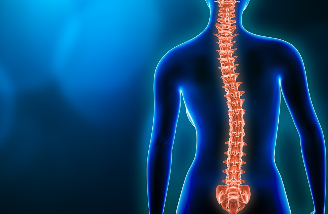
Tag Archives: registered physical therapists mississauga

Scoliosis: Types, Symptoms and Treatments

Scoliosis

Scoliosis

A side deviation or curvature in your spine is scoliosis. It could be in your neck, mid back and lower back. It can present in more than one area.

The most common area where it develops is mid-back. Cervical and lumber spine are very common  when it comes to two curvatures.

Type of scoliosis:

  • C-shape: This is most common type, there is only one-sided curve. The most common area for C shape to develop is in the thoracic spine and also lumber spine.  In this type, scoliosis side leg could be short compared to non-scoliotic side. Sometimes, there are  changes in the scapular level. Scoliotic side scapula will be lower than non-scoliotic side.
  • S- shape: In this type, two curvatures are present in the spine. One curvature is the primary scoliosis and the other curvature develops in compensation of the primary curvature. In this type, the scoliotic sided leg is short and the scapular level is higher than the non-scoliotic side. S-shape will bring other complications such as respiratory issues, cardiovascular issues, etc.
'S' Shaped curve and 'C' Shaped Curve

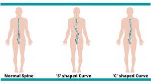
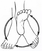
‘S’ Shaped curve and ‘C’ Shaped Curve

Causes of scoliosis:

Usually, the cause of scoliosis is unknown. However, following are a few possible causes mentioned:

  1. Poor Posture habits: People who have habits of not following ergonomics pattern of posture are at risk. One who has the habit of leaning and sitting mostly on their right or left, are also at risk.
  2. Carrying heavy stuff on back: People who have to deal with the delivery of heavy packets and lifting heavy objects are at risk.
  3. Family history: Sometimes, scoliosis runs in family. There are higher chances in these cases.
  4. Congenital limb length discrepancy: Congenital limb length discrepancy needs immediate correction otherwise; it will lead to scoliosis.
  5. Neurological disorder: Neurological disorders like cerebral palsy, multiple sclerosis, Down Syndrome which are progressive diseases have muscular imbalance.


Signs and symptoms:

  • Limb length discrepancy
  • Visible deviation of spine to one direction
  • Uneven shoulder and pelvis height
  • In anterior view, one sided rib is popping out.
  • Chronic neck and also lower back pain
  • Breathing difficulties in severe cases
  • Recurrent lung conditions like pneumonia, atelectasis, etc.
  • Pain and numbness
  • Fatigue due to muscle imbalance


Assessment of scoliosis:

Imaging: X-ray is the gold standard or MRI.

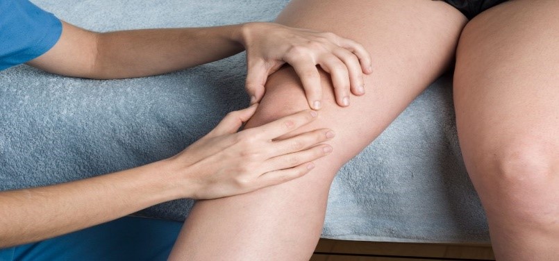
Physiotherapy Assessment:  assess in posterior view which means back view with open back.

  • Therapist will look for visible prominent border of scapula.
  • with bending forward the scapular border will be seen as well as deviation .
  • When leaning backward , a visible S shape or C shape will be seen.


Risk factors of scoliosis:

Scoliosis is bony disorientation which is irreversible. The best management is prevention. However, if it not managed well enough , it can lead to many complications.

  1. Lung disorders: Specially, thoracic spine scoliosis has a chance to affect the respiratory system as it disturbs the alignment of the rib cage. Deep breathing becomes limited or difficult and that is why the lower portion of the lungs get less air. As a result of this,  atelectasis develops in some areas of the lung.
  2. Limb length discrepancy: Lumber scoliosis has a major effect on pelvis alignment. As scoliosis is irreversible, it leads to permanent muscular imbalance. Thus, there is limb length discrepancy.
  3. Scapular winging: Thoracic scoliosis has drastic effect on scapular orientation.  The scapula takes part in shoulder movement. Indirectly, mid back, scapula and the shoulder joint are all affected because of spinal curvature.
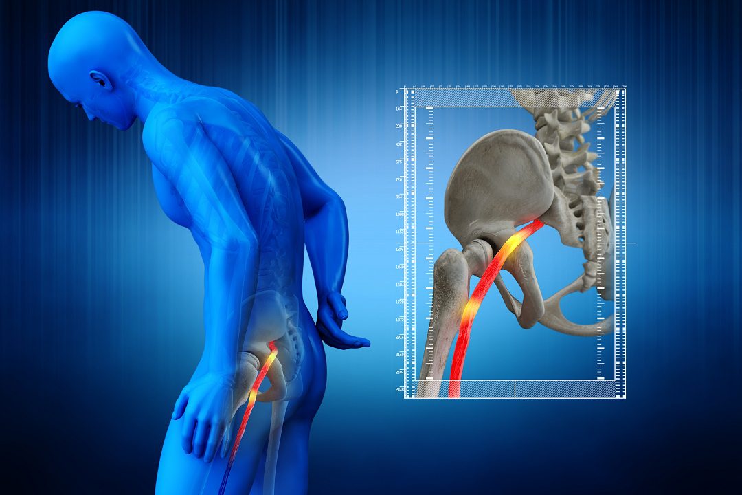
  4. Herniated disc in spine: Lumber spine scoliosis has a higher chance to develop a herniated disc. Therefore, you may develop pain in the lower back and down the leg  into the thigh, knee or ankle. Please seek immediate attention in this case.


Management of scoliosis:

 Scoliosis is a irreversible bony deformity.

  1. Posture: Contact your physiotherapist for a better understanding for well maintained posture at work and home.  Correction in pelvis alignment will help in preventing it from getting worse.
  2. Deep breathing exercise: Deep breathing exercise will help in maintaining lung function. Additionally, with breathing exercises, chest mobility exercises will help in maintaining flexibility in the spine and avoid lung conditions like pneumonia, atelectasis, etc.
  3. Spinal mobility exercises: Contact your physiotherapist for proper exercises. Your Physiotherapist can better assess scoliosis and based on that provide spinal mobility exercises to stop it from getting worse.
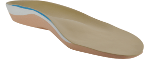
  4. Orthotics: orthotics adjust leg length discrepancy from scoliosis.
  5. Education: Finally, a physician or physiotherapist can help you to better assess and understand this condition.


Need some help?

Your concerns are best managed by a health care professional. Here at PhysioNow, we have many highly knowledgeable physiotherapists that can assist you with your issue. If you think you may have scoliosis or are diagnosed with it, book with PhysioNow today for your first assessment and treatment!

Chronic Ankle Instability Could Cause Your Ankle Not To Heal

Chronic Ankle Instability

Chronic Ankle Instability

Ankle sprains are one of the most common injuries. Rest, ice, compression and elevation, or the RICE rule, are the go-tos for ankle sprains. In most cases, the ankle will heal between 4-12 weeks. Many people will allow the ankle to heal on its own but a failure to seek treatment and physical rehabilitation for the ankle after just one injury is enough to leave it weaker and more vulnerable to future sprains. Which in turn could result in chronic ankle instability.

What causes ankle instability?

After an ankle injury, there is damage to the ligaments, tendons, and potentially even the ankle joint itself. Ligaments function to limit the normal movement in the ankle, and once they are injured, they may get lax (stretched out). Resulting in the ankle being more unstable. Additionally, the injury results in a loss of strength in the muscles that control the ankle movement, further increasing the instability of the ankle. Finally, it also results in a loss of sensory receptors in the ankle that communicate to our brain where our ankle is in space. This decreased sensation makes it harder to balance and control the ankle which contributes to the ankle instability.

How do I know if I have chronic ankle instability?

Questions to ask yourself include:

  • Does my ankle ever give out?
  • Do I avoid walking on uneven surfaces out of fear of falling?
  • Do I feel like I can’t “trust” the ankle?
  • Do I try to avoid putting all my weight onto one ankle?

If you said yes to these questions and you have had a severe ankle injury or a history of multiple ankle sprains, you may have chronic ankle instability.

Is it permanent?

Chronic ankle instability is not permanent it can improve with the help of a physical therapist and individualize exercise plan. An untreated ankle injury may be chronic when it is not given sufficient time to heal and is constantly being reaggravated. Thus, the first step to treatment will be a proper rehabilitation program with a health professional.

Who treats ankle instability?

Physical therapists are the first step in conservative management of chronic ankle instability. Your physical therapist will perform an assessment on your ankle range of motion, strength, and balance and will assess for any laxity, or more movement than is normal, within the ankle and ankle ligaments.

After an ankle injury, there is a loss in strength and proprioception (awareness of the position of the body in space). So exercises are given to re-strengthen the muscles, increase ankle stability, balance, and retrain the body awareness of the ankle. Furthermore, a brace and/or taping is recommended to support the stabilization of the ankle, especially for individuals who engage in high-risk activities like sports.

Chronic Ankle Instability

Chronic Ankle Instability Brace and Tapping

Chronic Ankle Instability Exercises

Chronic Ankle Instability Exercises

However, not all injuries are capable of recovering with conservative management. Thus, in very severe cases, surgery may be an option to repair the injured ligaments and give the ankle stability.

What happens if its goes untreated?

Without treatment, an individual will continue to have difficulties balancing, walking on uneven terrain, and putting weight onto the ankle. Subsequently, the instability may worsen over time and will have a much greater risk of reoccurring injuries to the ankle. Thus making it more challenging to recover from these injuries. Additionally, it can result in damage to the bones and cartilage within the ankle joint as the muscles weaken and fail to support the joint.  Therefore, to avoid severe injury it is highly recommended that individuals seek expert treatment.

If you are having stability problems with your ankle, book with PhysioNow today for an initial assessment and treatment. We have many capable physical therapists that will be able to help your ankle recover!

Trigger Finger

Trigger Finger? What can you do about it?

Trigger finger? What to do

Trigger Finger

Trigger Finger

Lately you’ve noticed that your finger seems to be clicking, and maybe it also catches or locks into a bent position. Sometimes you may need to use your other hand to help you “unlock” your finger. What is this? We call this condition trigger finger.

Your  finger locks in a flexed position. The position it locks in  resembles the position of a finger when it is pulling a trigger.

If you have trigger finger, you may also have:

  • Stiffness and swelling, especially in the mornings.
  • Painful or painless clicking, catching, locking into the bent position then popping straight.
  • Loss of full range of motion of the fingers into flexion and extension (bent and straight).
  • Nodule is felt with other hand.
  • Pain that radiates to the tip of the finger and/or into the palm.

Why does Trigger Finger happen?

An exact cause is unknown, it develops spontaneously or consequently to another disease, repetitive hand movements during a job, or recent trauma to the hand.

The condition occurs due to inflammation and narrowing in the “pulley” system in the hand. As seen in the image below, the tendons of our muscles must pass through a band of tissue in order to control the fingers. The tendon normally glides through this tunnel without issue but the tissue of the tunnel can thicken and narrow, combined with enlargement of or nodules developing in the tendon itself, resulting in impaired movement and the tissues blocking each other.

Trigger Finger Nodule and Tendon

How to treat trigger finger?

The first line of recommended treatment for any condition is conservative care. A physiotherapist can help you recover your hand function and quality of movement. These treatments
may include:

  • Soft tissue techniques, joint mobilizations, and stretches to try and increase the mobility in the
    area.
  • Modalities like ice, paraffin wax, and electrical stimulation to help with pain, inflammation, and swelling.
  • A home exercise program that focuses on increasing range of motion, strength, and gliding of the tendons
  • Education on wearing a splint (as shown below) which blocks the finger from moving excessively, allowing you to go about your daily life without aggravating the condition. Wear this between 6-10 weeks and your physiotherapist may ask you to wear them at night, during work, or as much as possible during the day.

What options do you have for treatment?

In some cases, this condition may be persistent and medical management with your primary provider. For example, options
may  include corticosteroid injections to reduce inflammation and the frequency of the finger locking. Unfortunately, patients may require further intervention when conservative treatment fails, in which case a surgical release of the finger may be indicated. When combined with physical therapy after the surgery, there is usually a rapid and significant improvement in function at relatively low risk. However, there are always increased risks to surgical procedures compared to conservative treatment and where possible, trigger finger should always be treated with non-surgical strategies first and surgery as last resort.

Need some help?

Let us  manage your trigger finger. Here at PhysioNow, we have many highly knowledgeable physiotherapists that can assist you with your issue. If you think you  have trigger finger or have been diagnosed with it, book with PhysioNow today for your first assessment and treatment!

Headaches? See how Physiotherapy can help you!

How do Headaches occur?

There are many different types of headaches with many different causes. Many people may blame it on the weather, stress, or work, and feel like it is out of their control and that nothing can help their headaches. If this sounds like you, there is treatment available and you do not have to live with your pain. Headaches that stem from your neck or jaw are treatable through physiotherapy and can result in a decreased intensity and frequency or even stop them completely. However, migraines can also be manageable with rehab.

What are headaches from the neck?

Cervicogenic Headache

Headaches that stem from the neck are known as cervicogenic headaches and are a type of referred pain. Referred pain means that the pain actually originates from another part of the body, and not the place where you actually feel the pain. Moreover, the head pain is actually coming from problems in the tissues and/or joints in the neck.

Common symptoms of cervicogenic headaches include:

  • Firstly, one-sided headaches
  • Second, neck stiffness
  • Decreased motion in the neck
  • Radiating pain to the eyes, shoulder, or arms
  • Lastly, they are triggered by certain neck positions or movements

What are headaches from the jaw?

TMJ Pain HeadacheHeadaches stemming from the jaw result from dysfunction in the jaw joint, anatomically known as the temporomandibular joint (TMJ). This joint connects your lower jaw to the skull and is used in actions such as talking and chewing as it allows the opening and closing of the mouth. Therefore, headaches are one of the common symptoms of jaw dysfunction. Though they are lesser known, it is commonly missed as a source.

Common symptoms of TMJ dysfunction include:

  • Clicking on the jaw
  • Pain or tenderness with jaw movements (biting, talking, chewing, etc)
  • Headaches, especially in the morning (due to grinding and clenching of the jaw during the night)
  • Also, neck stiffness

How can physiotherapy help?

Your physiotherapist will provide a comprehensive assessment of your neck and jaw to determine a specific treatment plan for you. The evaluation may include observing your resting posture, and the way your jaw and neck move. Also,  how much movement is available in your jaw and neck, and more.

Your treatment plan may include strengthening weak muscles, correcting posture and faulty movement patterns, stretching and mobility exercises, manual therapy techniques like soft tissue massage and joint mobilizations to reduce pain and increase range of motion. They may use modalities like heat, electrotherapy and acupuncture as well depending on your condition and goals of treatment. Your physiotherapist will work closely with you to develop an individualized plan of treatment including a home exercise program, and education on self-management of your condition at home.  They can also inform you and help you get in contact with other healthcare practitioners that may need to be involved in your care.

If you are experiencing persistent headaches, book with PhysioNow today for your first assessment and treatment!

Say Goodbye to Knee Pain with Orthopedic Custom Knee Brace!

Say Goodbye and Beat knee pain with an Orthopedic Custom Knee Brace!

Here at PhysioNow, we can help you recover from your knee pain faster by providing you with a Custom Knee Brace. There are many types of knee braces available for purchase. Therefore, it can be overwhelming to know which brace is best for you so does the type of knee pain. In this article, we will give you an overview of custom knee braces. Also, provide instructions on how to start the process of ordering your custom knee brace.

Who can get a custom knee brace for knee pain?

Custom knee braces are most used for individuals with/after:

  • Post-operative recovery
  • Knee instability
  • Osteoarthritis
  • Ligament or meniscal injuries

Moreover, the brace is constructed using precise measurements taken from your leg. It is designed to only fit you.

Why get a custom knee brace? Is it worth it for knee pain?

Many patients have found that bracing has returned them to a level of function that they were previously thought to be unattainable after their injury. Some of the benefits of a custom knee brace include:

  • Firstly, get you back to doing the activities you love.
  • Second, reduce pain and increase the function of the knee.
  • Support and prevent further injury while you await imaging, surgery, or after surgery.
  • Individualized to your needs and level of activity ex. Lifestyle vs performance braces.
  • Ideal for long-term use.
  • Lastly, lightweight and can fit underpants for a subtle look.

What types of custom knee braces are there?

The most common type of custom-made knee brace is a rigid knee brace. They are usually made of metal, with a hinge to allow the knee to bend, and are the most supportive type of brace.

They are designed for individuals who need moderate-maximum support. The conditions that require a rigid custom knee brace include moderate to severe osteoarthritis, and athletes that need more support after an injury. Also, pre and post-surgical repair of ACL, MCL, PCL ligaments, or menisci.

Moreover, under the umbrella of rigid knee braces, there are unloader braces for osteoarthritis. These braces provide more support to the painful, arthritic side of the knee to offload it. This can preserve the joint space while reducing knee pain and increasing knee function.

Another category of rigid knee braces is ligament braces for ACL, MCL/LCL, and PCL injuries, including pre and post-surgical repair. These provide knee support to the sides of the knee to prevent any unnatural twisting or movements. However, our PhysioNow team of physiotherapists will be able to determine which brace is best suited to your goals and lifestyle.

How can I get a custom knee brace?

Follow these simple steps below to get one.

  1. Contact us at PhysioNow to book an appointment time for a custom knee brace assessment.
  2. On the day of your appointment, your PhysioNow Brace Specialist will perform a series of tests and ask you questions to determine which type of brace is right for you.
  3. Make sure you tell your Physiotherapist what activities you are currently doing, and any activities that you would like to get back to doing (ex. Golf, running, walking)
  4. Your PhysioNow Brace Specialist will measure and take recordings of your knee, and discuss any considerations like the desired weight of the brace, special features (ex. Straps and clips), materials, colors, etc
  5. Lastly, we will be able to make inquiries to determine whether your custom knee brace can be covered by extended health care coverage. Care plans that include physiotherapy service coverage will usually have coverage for a custom knee brace as well. This process requires a physician’s note, but our clinic will be able to walk you through the process and provide you with the documentation needed to send to your insurance company for inquiry.

Once ordered, delivery will normally take around 7 business days but can be expedited upon request. We will ensure that the brace fits properly and can make adjustments if needed once delivered. So get your own customized brace NOW!