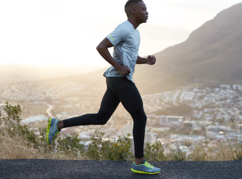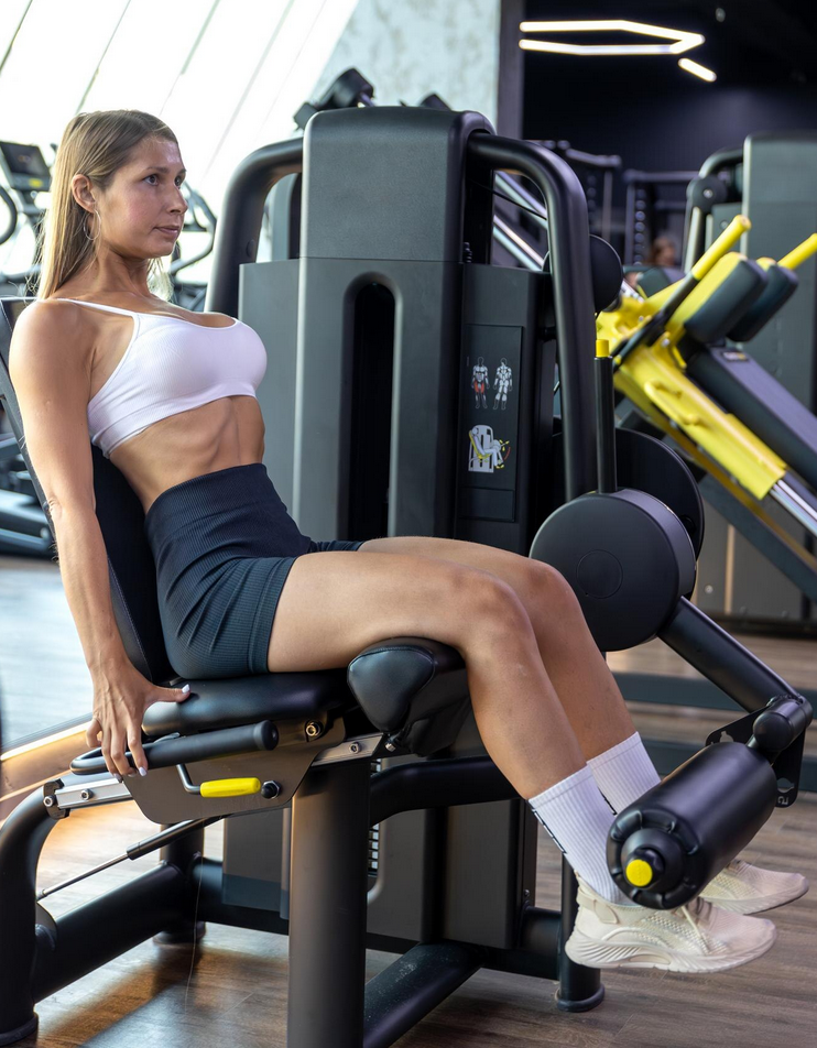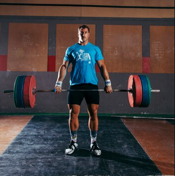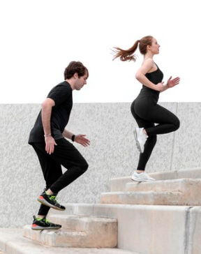

With the World Cup in full swing, the soccer season is in full swing. Even if you’re not a professional athlete, whether you’re playing in a league, drop in, or registering for a friendly tournament, you should always be on the lookout for overuse injuries. Most of us are familiar with that bone-deep fatigue after the final whistle blows or the timer hits 0, and the adrenaline comes to the end. It’s that legs shaking feeling, a pain that you didn’t notice or were able to ignore before that we call a post-tournament or post-game fatigue. In this blog, we’ll break down why your body feels this way, what it means to you, and how to take proper care of yourself so you don’t have to watch from the sidelines with an injury.
What is post-tournament fatigue?
Tournaments require a lot from your body, asking you to play at your peak several times in quick succession. This leaves your body without a lot of rest time to repair, resync, and replenish. That drained state happens because of:
- Nutrient depletion: Playing requires a lot of fuel, which comes from calories. Our body’s main source of fuel is stored carbohydrates and over the course of an intense series of games, the body’s storage can be depleted
- Muscle micro-tears: High intensity movements like sprinting, kicking, and jumping sends a lot of force through our muscles, causing micro tears. These micro tears are a normal part of training and how the body learns to adapt and build itself stronger. The issue comes when there is a huge increase in the volume of play, such as during a tournament. As your muscles accumulate all this damage without sufficient time to repair itself, they get less efficient and the risk of injury increases.
- Central nervous system fatigue: Just like our muscles, our nervous system can actually fatigue as well. Research has shown that the fatigue is associated with the neurotransmitters (chemical messengers) in the brain. Ultimately, the result is reduced physical performance and even brain fog.
What are overuse injuries?
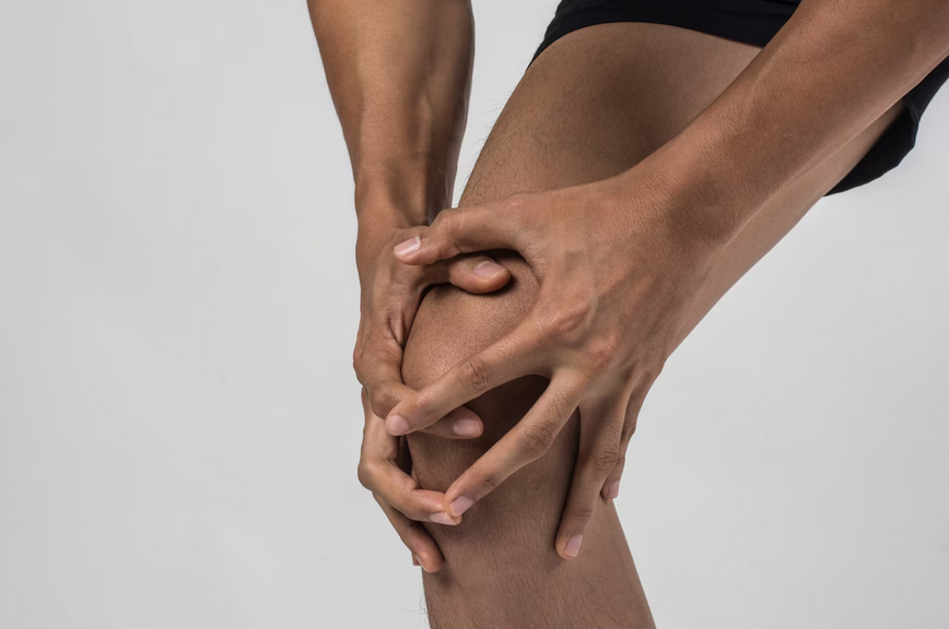
Unlike an acute injury like a muscle or ligament strain, overuse injuries sneak up on you over time. They may start as just a mild nagging pain that comes and goes, then gradually becomes more frequent or more intense over time. If not treated early on, they can develop into bigger issues that affect your daily life and ability to play. Another name given to overuse injuries is repetitive stress injuries.
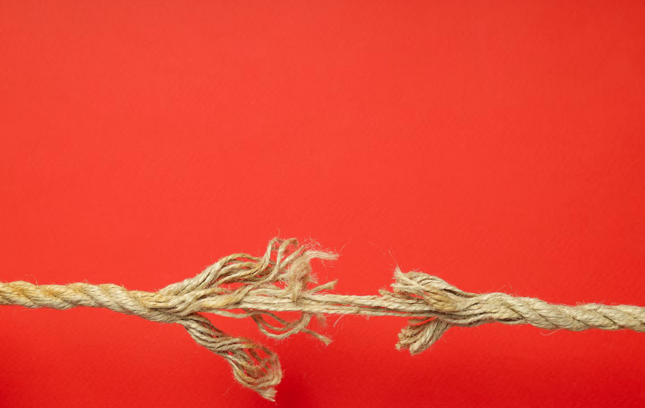
An easy way to think about it is that the demand (what you’re asking the tissue to perform) is overloading the supply (what your tissue is capable of). Like a rope that is supporting too much weight, it starts to fray and break. Thankfully, with the right training, overuse injuries can be caught early on and prevented.
How do I spot an overuse injury?
Overuse injuries tend to follow similar patterns even if the body parts can be very different:
- Timeline of pain: Some muscle soreness, known as DOMS (delayed onset muscle soreness) is normal 24-48 hours after exercise. If there has been a recurrent or consistently present ache, stiffness, or discomfort, it is likely the start of an overuse injury
- Warm-up pain: Usually worse after waking up, sitting for a long time, or first starting a workout. After moving around for a bit the pain goes quickly goes away, then may return worse in a couple of hours
- Pinpoint pain: In a lot of tendon conditions, people are able to directly point towards the source of their pain. When that spot is pressed, it recreates their pain
- Compensatory movements: If you’ve noticed that your running gait, the way you swing your racquet, or your overall performance has decreased, that could be a sign of general fatigue or an overuse injury in the making
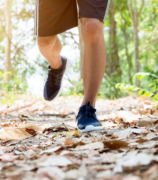
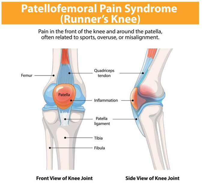
What are the most common soccer overuse injuries?
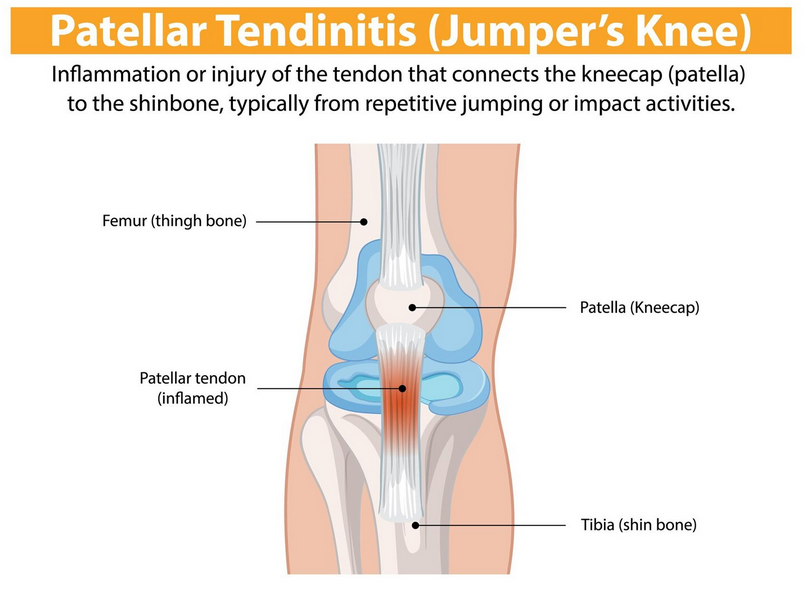
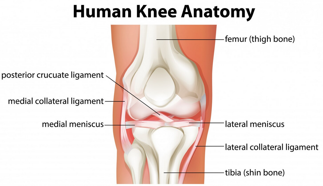
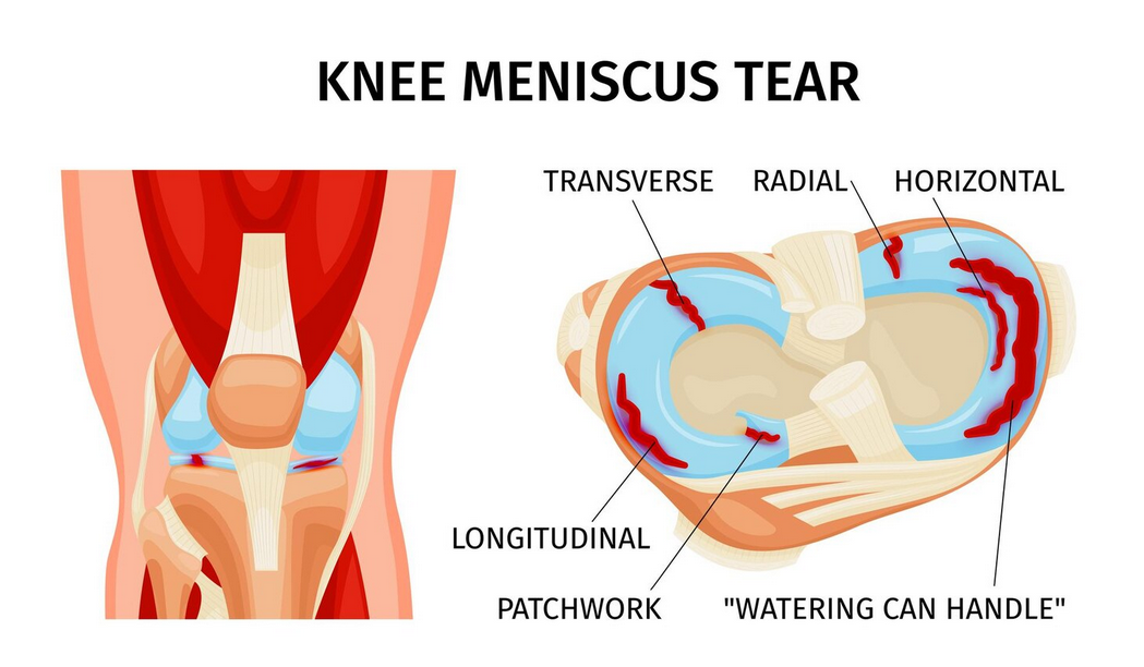
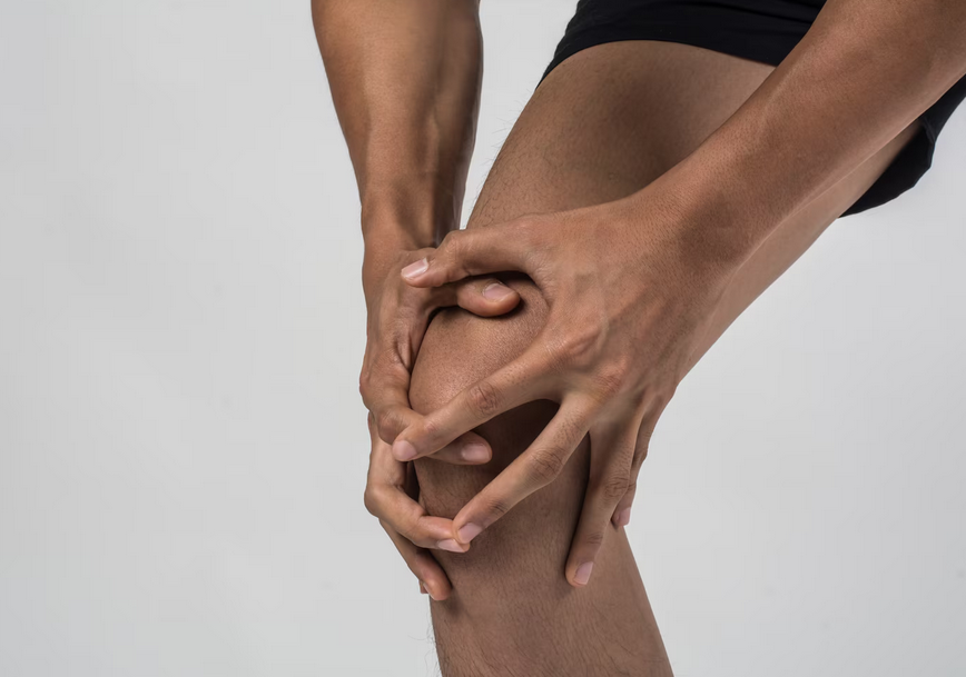
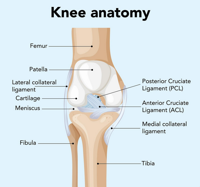
- Patellar tendinopathy: Also known as Jumper’s Knee. This is an overuse injury of the patellar tendon right below the knee cap. Constant sprinting, stopping, jumping puts a lot of pressure on this tendon. Most people will have a warm up pain and find it hard going down stairs
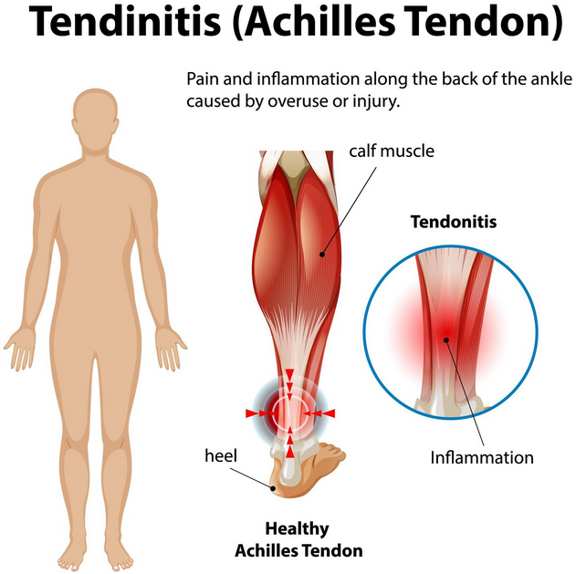
- Achilles tendinopathy: The Achilles tendon is one of the strongest tendons in our body that helps us to generate and accept force through our ankles and feet. Most people complain about pain when doing anything high impact such as jumping or jogging
- Shin splints: Your shin bone (tibia) and the surrounding musculature can become inflamed from repeated high impact stress. If left untreated, shin splints can turn into stress fractures.
How should I take care of myself after a tournament?
The immediate 48 hours after should be relatively restful. Light gentle active recovery such as walking and stretching are great to help blood flow and recovery, without contributing excess pressure to already compromised tissues. Lots of sleep, hydration, nutrition and sleep is essential during this stage.
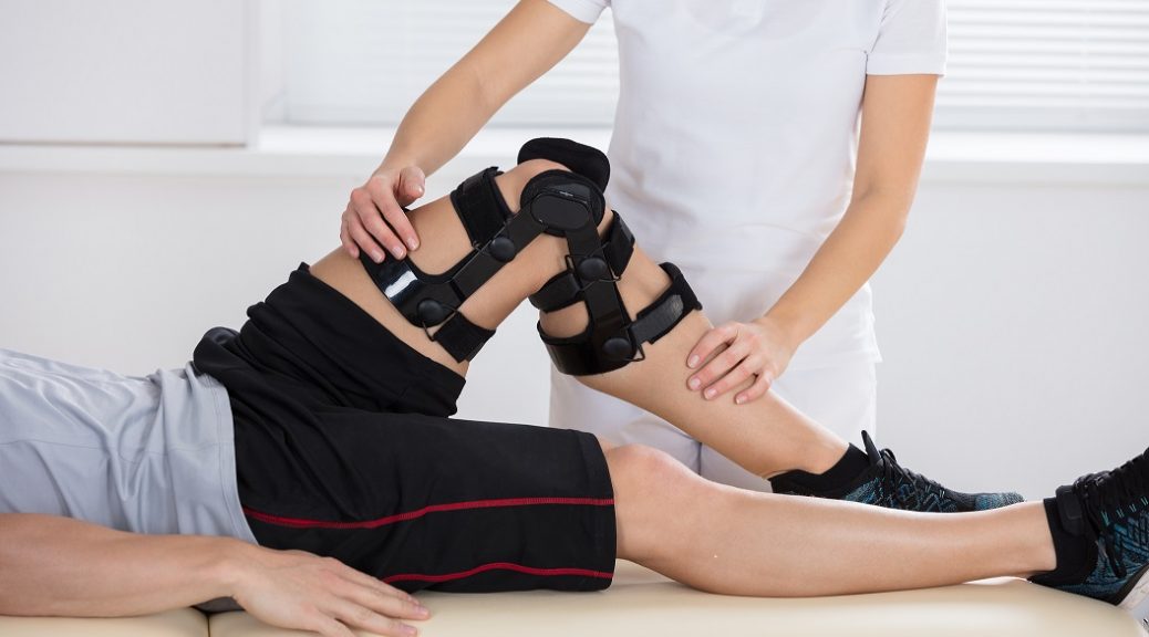
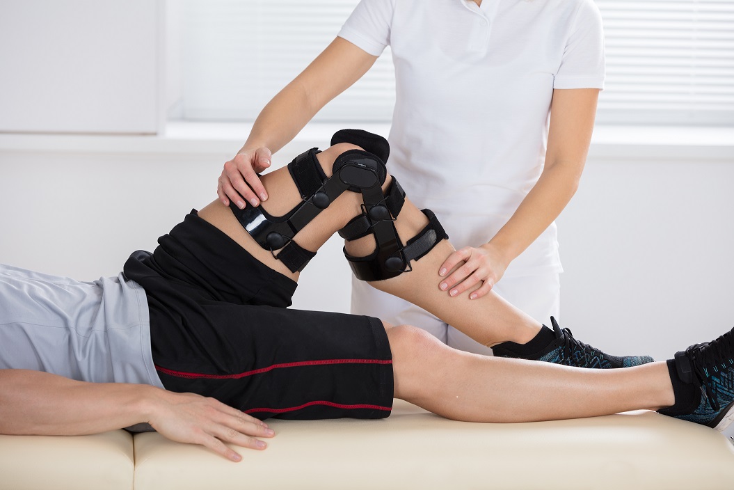
Afterwards, if there are any pains lasting or sharp pains present, a rehabilitation plan should be considered with Physiotherapy to address them. Overuse injuries are always easiest to heal the earlier you address them. When you let them progress, you run the risk of it becoming a more serious issue and might even have to take time off from your sport. Pushing through is not always the solution.
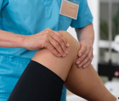
A Physiotherapist can help you identify any weak points in your training, muscle compensations, faulty movement patterns, and more. They will use various treatment methods including manual therapy, therapeutic exercises, modalities like shockwave therapy and more depending on your unique circumstances.
Dealing with a stubborn overuse injury?
Don’t wait any longer, come to PhysioNow to get started on your recovery. Summer is flying by fast and we want you to make the most of your soccer season and stay away from any other sports injuries. We have 10 locations across the West GTA including Burlington, Oakville, Mississauga, and Etobicoke. Book with PhysioNow today for your first assessment and treatment! Or contact us at Email: applewood@physionow.ca or Phone: 289-724-0448 for more information.