Tag Archives: Sports Injuries

De Quervain’s Disease

De Quervain’s Disease/ Texting thumb.

De Quervain's Disease

De Quervain’s Disease

De Quervain’s Disease or nowadays known as texting thumb is a painful inflammation of tendons in the thumb that extend to the wrist. The rubbing of the inflamed tendon against the canal it passes through causes pain at the base of the thumb and into the lower arm. It is commonly seen in females over 40 years of age.

Causes of De Quervain’s Disease

1. Simple strain injury to the tendon.
2. Repetitive motion injury. Workers who perform rapid repetitive activities involving pinching, grasping, pulling or pushing are at increased risk. Specific activities including intensive mousing, trackball use, and typing. Other activities including bowling, golf, fly-fishing, piano-playing, sewing, and knitting can also cause De Quervain’s Disease.
3. Frequent causes of De Quervain’s Disease include stresses such as lifting young children into car seats, lifting heavy grocery bags by the loops, and lifting gardening pots .
4. De Quervain’s Disease often occurs during and after pregnancy. Factors may include hormonal changes, fluid retention and more lifting.
5. Rheumatoid arthritis.

De Quervain's Disease

De Quervain’s Disease/ Texting Thumb

Onset and Symptoms of De Quervain’s Disease

Onset can be gradual or sudden. Pain is felt along the back of the thumb. There can be Pain directly over the thumb tendons, and pain may travel into the thumb or up the forearm. The bottom of the thumb or the side of the wrist might also be sore or swollen.
It may be hard and painful. Symptoms may get worse when the thumb is moved, particularly when pinching or grasping things. Some people also have swelling and pain on the side of the wrist at the base of the thumb. The back of the thumb and index finger may also feel numb. People might experience a funny sound like a squeak, crackle, snap, or creak when they move the wrist or thumb.
If the condition is not well addressed, the pain can spread up your forearm or down into your thumb.

How is De Quervain’s Disease diagnosed?

De Quervain’s Disease is diagnosed based on history and physical examination. X-rays, or ultrasound may be used to rule out other causes of pain.The Physiotherapist may use special tests to help diagnose De Quervain’s. More information can be found here.

Physiotherapy Treatment

Your Physiotherapist will likely recommend that you wear a specific wrist splint with a thumb spica for 4 to 6 weeks . PhysioNow carries these wrist splints in stock. You’ll also need to stop doing activities that worsen the condition.

Wrist brace with thumb spica

De Quervain’s Disease

The physiotherapist after a thorough assessment could choose different therapy approaches to help with the swelling, pain, and function.
These treatments may include Ultrasound, K-taping, acupuncture, and manual therapy. Other Physiotherapy treatments may include specific exercises focusing on range of motion, strength, and flexibility. These would be given for a safe and effective return back to function.

Recovery times vary depending on your age, general health, and how long you’ve had the symptoms.
If your disease has developed gradually, it’s often tougher to treat. So, it may take you longer to get relief. Your doctor may give you anti-inflammatory medication, or may inject the area with steroids to curb pain and swelling.

In our experience at PhysioNow, more than 99% of people with De Quervain’s Disease get better with Physiotherapy treatment provided. If however, you are one of the outliers, your doctor may recommend surgery. The operation would release the tendon’s tight covering so that the tendon could move smoothly. It’s an outpatient procedure, which means you go home afterward. Your doctor will recommend physiotherapy after surgery which includes an exercise program to strengthen your thumb and wrist.

If you or someone you know suffers from De Quervain’s Disease, please call us today. Our skilled Physiotherapists can Help!

Trochantric bursitis


Trochanteric bursitis


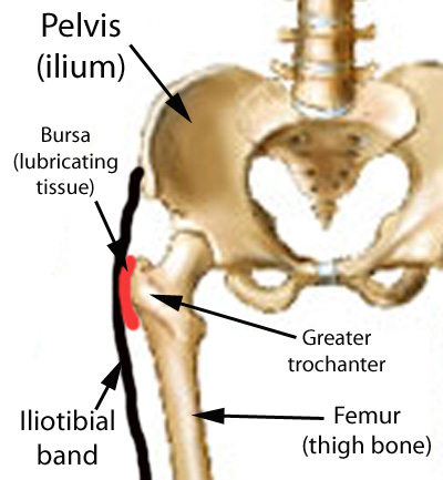
The Trochanteric Bursa is a fluid filled sac near the hip joint. It cushions between the bones and the muscle and acts as a shock absorber. Trochanteric bursitis is the inflammation of the bursa which is at the outside of the hip.

When this bursa is irritated it causes pain on the outside of the hip and thigh.  Sometimes you may feel the pain down to the knee on the same side.  Another symptom is pain which will stop you from lying on your painful side.  It will be painful when you press on a specific spot on the outside of the hip.  The pain gets worse when climbing stairs, getting up from a low chair getting out of a car.  Sitting with crossed legs hurts.  Both  walking and running may also cause increased pain.

Causes of Trochanteric Bursitis

  • Any direct Injury to the outside of the hip like falling, or bumping into an object.
  • Incorrect posture due to muscle imbalance
  • Leg length difference.
  • Stress of soft tissues surrounding the hip due to hip Osteoarthritis
  • Friction of the ITB (Ilio Tibial Band: a muscle along the side of the hip)
  • Overuse injuries like standing for long periods of time
  • Other diseases which affect the hip like Rheumatoid Arthritis
  • Previous surgeries around the hip
  • Very rarely infection of the bursa

Registered Physiotherapy Treatment

Your Registered Physiotherapist can help you to reduce the pain and inflammation.  They will use electrotherapy modalities, taping, or acupuncture.  They will give you  advice on activity limitations in the early stages.

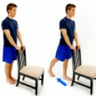
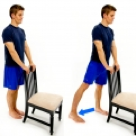
Later the treatment is focussed towards increasing the flexibility and strength in the muscles.  Your Physiotherapist will  increase the hip joint range of movement and correct your hip joint mechanics.

Trochanteric bursitis

Trochanteric bursitisTrochanteric bursitis

Finally the focus is towards preventing it from happening again.

General Advice on prevention

Your Registered Physiotherapist will look at your posture, hip stability, and core strength.  They will look to see if you  need Orthotics to address any problems with your legs or feet.

 Tips for prevention of Trochanteric Bursitis:

  • Whenever you start a new activity, try to take it slow and build up your activity level gradually.
  • Start with  less force or resistance, less repetitions and progress gradually.
  • Avoid or modify repetitive activities if it is putting any stress on the hip joint
  • Your footwear should be comfortable
  • Maintain flexibility and strength in your lower extremity muscles
  • Improve your core muscle strength

If you or someone you know may have Trochanteric bursitis, call PhysioNow Today!  We would be happy to get you started on your treatment and back to enjoying all the things you love to do!


Vertigo

Benign paroxysmal positional vertigo(BPPV)

Vertigo

Vestibular Systems
• Comprises five sensory organs that provide your brain with information about head position and movements including head rotation, linear movements and static positions of the head relative to gravity
• Five sensory organs including 3 semicircular canals and 2 otoliths


BPPV is a mechanical problem in the inner ear. It occurs when some of the  crystals that are normally embaded in gel in the ear become dislodged.  They then can move into one or more of the 3-fluid filled semicircular canals.

Benign paroxysmal positional vertigo(BPPV) is  one  of the most common causes of vertigo. It creates a false sensation of spinning.
• Benign: it is not life threatening
• Paroxysmal: it comes in sudden, brief spells
• Positional: it gets triggered by certain head positions or movements
• Vertigo: a false sensation of rotational movement


Symptoms
a)      Dizziness
b)      Vertigo (sensation of spinning)
c)       Nausea
d)      Sense of imbalance or unsteadiness
e)      Poor gaze stability
f)       Vision disturbance

Visual Coordination screening


Diagnosis

• The relationship between the inner ears and the eye muscles are what normally allows us to stay focused on our environment while the head is moving.  The dislodged crystals make the brain think you are moving when  you are not. This mistakenly causes the eyes to move, which makes it look like the room is spinning. This is called Nystagmus.

• The Nystagmus will have different characteristics that allow the practitioner to identify which ear the displaced crystals are in and which canal is involved.
• The most common tests are DIX hall pike and Roll test.
• There are two types of  BBPV.  One type, where the loose crystals can move freely in the fluid of the canal(canalithiasis). The more rare type is one where the crystals are thought to be hung up on the bundle of nerves that sense the fluid movement(cupulolithiasis).

Physiotherapy Treatment
• One maneuver that is used for the most common location and type of BPPV is called the Epley maneuver. However , that will not work for all people . Often people have tried the Epley maneuver themselves or had it performed on them without success.
• In the vast majority cases, BPPV can be corrected mechanically by a Registered Physiotherapist. Once your  provider knows which canal is involved and what type it is , we can take you through the appropriate treatment maneuver.

Check out this video for an example of the  Epley Maneuver.  Please do not attempt this yourself until you have been properly assessed and screened by one of our Registered Physiotherapists to see if this maneuver will be appropriate for your condition!

If you have any further questions, please call PhysioNow today! Our experienced Vestibular Physiotherapists would be happy to help you.

Baseline Testing Concussions

Baseline Testing

Why is baseline testing Concussions important?

There are no  special tests to diagnose and gauge the severity of concussions.  Athletes are recommended to undergo baseline testing before the beginning of the sports season. Baseline test results are helpful  for most healthcare professionals  involved in an athlete’s concussion care. This includes Physiotherapists, Physicians, Concussion Specialists, and Vision Therapists.  Baseline tests let the Physiotherapist compare the before injury  function to the results of post concussion baseline testing. It’s a great tool  to measure the severity of concussions.  It helps therapists to make  treatment goals.  Safe return to sports following concussions can be determined with this tool.


Components of Baseline testing  Concussions


  • Impact Test, This is the gold standard test.  It  looks at different functions of your brain before and after concussion. It is a computer based test. It usually takes about 30 minutes. The test assesses your ability to process information, and remember things.  It also tests how quickly you can follow  instructions/tasks. It’s the most scientifically validated test and recently got approved by the FDA.


  • Visual Coordination screening 
    Visual Coordination screening
     Following concussion, functioning of the visual system  is greatly affected. This involves the eyes and part of the brain that processes visual information.   Correct vision and clarity of  vision is not affected. However, the ability of your eyes to move together is affected. This usually leads to symptoms like dizziness, difficulty focussing,  and headaches. Testing the visual system before the sports season provides valuable information about the function of the visual system.  It allows for treatment of any problems.


  • Balance and Postural stability– With Post-concussive injury, both static and dynamic balance is affected. You can feel dizzy with standing, change in position or with certain movements. Most sports require an intact balance system to improve performance.


Registered Physiotherapists at PhysioNow are trained to conduct baseline testing before the beginning of  the sports season and following concussive injury. Registered Physiotherapists at Mississauga are skilled to assess any deficits in your baseline testing and treat them. Visual coordination exercises  will be done to help you  return to sports faster .

Please call today to get your baseline testing done or to treat your concussion.

Foot Pain Treatment Mississauga

strong>Foot Pain Treatment Mississauga
Trending Now: Blue Jays’ Jose Batista on 15 day disabled list

Foot Pain Treatment Mississauga

Blue Jays’ Jose Batista hurt his big toe during a June 16 game against Philadelphia. After running into the outfield wall, Bautista had to leave the game early because of pain. An MRI showed that he had suffered a sprain of his left big toe. He had to be in a walking boot for the time being. It may seem like it’s an excessive treatment plan for just a simple sprain. In reality sprains can sometimes be more complicated than a fracture.

What is a Ligament?

A ligament is a soft tissue structure that connects two bones together. Ligaments do not receive a good blood supply like muscles or tendons and as a result, they cannot heal as well.

Types of Sprains

A sprain is defined as a stretch or tear of a ligament.
Furthermore, sprains can be classified into three groups based on the severity of injury. A grade I sprain means that there is mild damage to the ligament and the joint is stable. A grade II sprain means that there is a partial tear in the ligament and the joint is overall less stable or loose. The most severe type of sprain is classified as grade III and it means that the ligament has completely torn or ruptured and the joint has lost stability.

How can a Registered Physiotherapist help with Foot Pain treatment Mississauga?

A Registered Physiotherapist helps with all three types of sprains. The treatment for sprains starts with rest and reducing inflammation. Depending on the type of sprain, it may be necessary for immobilization in order for this to occur. For example a grade II sprain you can get a removable walking boot. A grade III sprain you can get a cast. Once inflammation is reduced and pain is better, treatment seeks to improve range of motion and strength and a return to activity or sport.

Since Jose Batista has been wearing a walking boot, it seems that his injury is at least a grade II. Considering the poor blood supply of ligaments and the severity of his particular sprain it is definitely best for him to be out of the game for at least 2 weeks. Proper foot pain treatment Mississauga in the early phase of injury is very important so that the ligament is well protected and given the best chance for healing. Once the walking boot comes off, his injury will be re-evaluated and the Blue Jays’ medical team will decide on his further treatment plan and eventual return to play.

Jose Bautista’s toe injury improving

If you have been injured, call PhysioNow today! They can help with Foot Pain treatment Mississauga!