Tag Archives: physiotherapy

Achilles tendonitis Shockwave therapy

Stubborn Pain? Try Our New Shockwave Therapy!

Shockwave therapy

What is shockwave therapy?

Shockwave therapy is also known as extracorporeal shockwave therapy. It is a treatment which uses low energy acoustic wave pulsations. The modalities can be directly applied to the area of injury using a gel as a medium. Lately, it is becoming one of the common treatment modalities seen in physiotherapy clinics.

Why is shockwave therapy used?

Importantly, shockwave can be beneficial in treating many musculoskeletal conditions, especially chronic ones. Firstly, it is used to stimulate the body’s natural healing process. Additionally, it can help you to decrease your pain levels by stimulating the nerves around an injured area. Also, the therapy increases blood circulation to the area and can also accelerate protein synthesis, cell growth, and break down stubborn calcium deposits in tissues.

How does shockwave therapy work?

The machine generates shockwaves, which is mechanical energy, throughout the tissue. Consequently, this stimulates a chain reaction or response in the cells which promotes tissue regeneration. The mechanism of action is quite complex as it happens at the cellular level but overall, results in a positive healing response and pain relief effect for the body. The shockwave therapy machine includes different interchangeable heads with different penetration capacity.

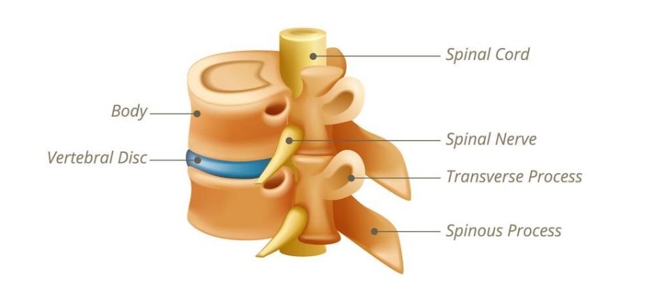
What type of injuries can be treated?

Shockwave therapy applied to a patient's heel

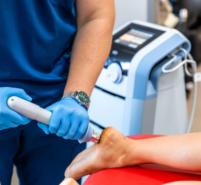
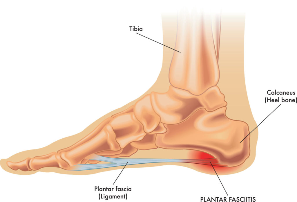
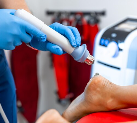
The foot is one of the most commonly treated areas with shockwave therapy

Shockwave was initially used to disintegrate renal stones in the 1980s. From there, it was discovered to be effective at treating bone spurs and calcified tissues. The following are a list of injuries that are most commonly treated by shockwave. Even if your injury is not on this list, shockwave may still be an option for you!

  • Planter fasciitis
  • Patellar tendinopathy
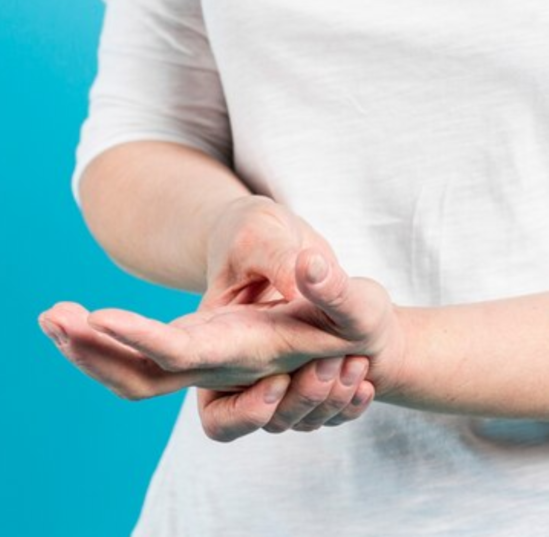
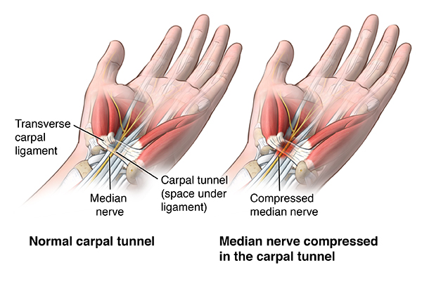
  • Upper extremities tendinopathy
  • Lower extremities tendinopathy
  • Tennis elbow
  • Golfers elbow
  • Hamstring injuries
  • Achilles tendinopathy
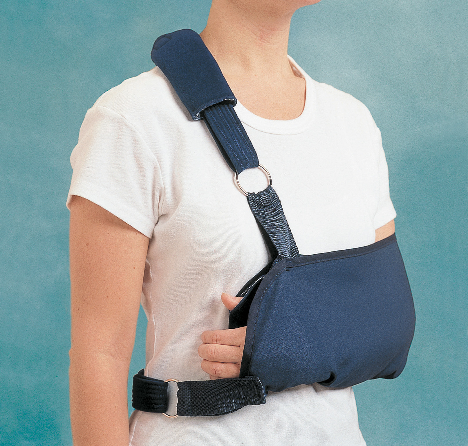
  • Rotator cuff injuries
  • Sports injuries

When to avoid it:

Shockwave therapy should not be used if you have the following conditions:

  • Active cancer
  • Pregnancy
  • Open wound
  • Infection
  • Pacemaker
  • Blood clotting disorders

Is it painful?

Usually, most patient do not find it painful, but they may feel some kind of discomfort during the application of it. Discomfort also tends to vary based on the area of treatment, with some being more sensitive than others. After the treatment, patient might feel some soreness and notice some redness around the treatment area. However, this is perfectly normal and will go away within a couple of days.

What does a treatment look like?

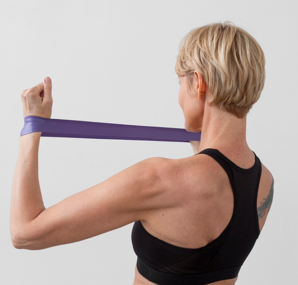
During your visit the physiotherapist will check your appropriateness for shockwave therapy. Firstly, they will conduct an assessment to rule out any contraindications and precautions for your injury and other overlapping conditions. The one session for therapy is usually between 10-15 mins, depending on the area and condition. Oftentimes, patient feels the difference in their symptoms with just a few sessions, some even immediately after treatment! Usually, your therapist will start with lower intensities and will gradually increase it with respect to your pain tolerance. Our goal is to ensure each patient is comfortable and knows what to expect with treatment. Additionally, shock wave therapy is used alongside other physiotherapy treatments like manual therapy and therapeutic exercises to get the best results.

Need help or more information?

If you are interested in receiving shockwave therapy or want to learn more about it, please contact us at PhysioNow. Fortunately, we have many experienced physiotherapists across Burlington, Oakville, Mississauga and Etobicoke who would love to answer your questions. Currently, shockwave therapy is available at our Applewood location located just off the QEW at the Cawthra exit!

Fractured Bone? 5 Things You Should Know

Being diagnosed with a fracture can be a challenging experience, but proper care and attention to your recovery can significantly impact the healing process. Here are five important things to do after being diagnosed with a fracture:

1. Follow Medical and Surgical Advice:

fractured bone types

Types of fractured bones

Listen carefully to your healthcare provider’s recommendations and follow their prescribed treatment plan. This may include wearing a cast or brace for a set number of weeks, taking medications as prescribed, and attending scheduled follow-up appointments. Additionally, if you had a surgical repair, your surgeon may have additional instructions for you to follow. Compliance with this advice will ensure that there are minimal interruptions to the healing stage, and any issues can get caught and managed quickly.

2. Rest and Protect the Fractured Area:

Give your body the time it needs to heal by resting the affected limb or body part. For example, avoid putting excessive weight, movements, or stress on the fractured area. Follow any mobility restrictions to prevent further injury and support the healing process. The most common ways these instructions are communicated are as following:

    1. Non-weightbearing (NWB): No weight is allowed on the limb at all.
    2. Toe-touch weightbearing (TTWB): Only the lightest touch is allowed for balance.
    3. Partial weightbearing (PWB): Usually given as a percentage like 25, 50, or 75%.  This indicates how much of your full weight you can put on the area.
    4. Weightbearing as tolerated (WBAT): You can weight bear as much as it feels comfortable, your limitation will be based more on your pain and/or fatigue.
    5. Full weightbearing (FWB): You can use your full body weight, there are no restrictions.

3. Use Supportive Devices:

Crutches used after a fractured bone

Crutches used after a fractured bone

If your healthcare provider recommends assistive devices such as crutches, a sling, or a walking boot, use them as instructed. These devices are designed to help you move safely while minimizing stress on the fractured bone. Proper use can prevent complications and promote healing.

4. Monitor for Complications:

Keep an eye out for potential complications, such as signs of infection (increased redness, swelling, warmth, or discharge at the fracture site) or circulation problems (numbness, tingling, or changes in skin color). Report any concerns to your healthcare provider immediately.

5.Engage in Rehabilitation and Physical Therapy

Once your healthcare provider gives the go ahead, engage in rehabilitation and physical therapy. These activities are designed to improve joint mobility, strengthen muscles, and enhance overall function. Rehabilitation plays a crucial role in restoring normal movement and preventing long-term complications from your fractured bone.

Overall, it’s important to maintain open communication with your healthcare provider throughout the recovery process. Additionally, report any unusual symptoms, such as increased pain, swelling, or changes in sensation, promptly. Most of the time, follow-up appointments and imaging studies may be necessary to monitor the progress of your bone’s healing.

Recovering from a fracture takes time, and patience is key. It’s essential to prioritize the injury, adhere to medical guidance, and seek support from healthcare professionals to ensure the smoothest recovery. If you have recently experienced a fracture and are looking to start your rehabilitation, PhysioNow has many experienced clinicians to assit you. Book with PhysioNow today for your first assessment and treatment!

Osteoarthritis: How Physiotherapy Can Stop Your Pain

Osteoarthritis in the knee. Image by jcomp on Freepik

What is osteoarthritis (OA)?

Osteoarthritis (OA) is the most common form of arthritis. Alternatively, some people call it degenerative joint disease or “wear and tear” arthritis. It occurs most frequently in the hands, hips, and knees.

With OA, the cartilage within a joint begins to break down and the underlying bone begins to change. These changes usually develop slowly and get worse over time. Eventually, it can cause pain, stiffness, and swelling. In more severe cases, it also causes reduced function and disability; some people are no longer able to do daily tasks or work. Osteoarthritis (OA) is the most prevalent joint disease and a leading cause of disability in older adults.

Signs and symptoms of osteoarthritis:

  • Pain or aching
  • Stiffness
    Decreased range of motion (or flexibility)
  • Swelling

Risk factors:

  • Joint injury or overuse—Injury or overuse, such as repetitive knee bending or stress, can cause damage and increase the risk of OA in that joint
  • Age—The risk of developing OA increases with age.
  • Gender—Women are more likely to develop OA than men, especially after age 50
  • Obesity—Extra weight puts more stress on joints, particularly weight-bearing joints like the hips and knees. Consequently, this extra stress increases the risk of OA in that joint. Obesity may also have metabolic effects that increase the risk of OA
  • Genetics—People who have family members with OA are more likely to develop it. Additionally, people who have hand OA are more likely to develop knee OA
  • Race— Some Asian populations have lower risk for OA.

How is it diagnosed?

A doctor diagnoses OA through a review of symptoms, physical examination, X-rays, and lab tests. Furthermore, a rheumatologist, a doctor who specializes in arthritis and other related conditions, can help if there are any questions about the diagnosis.

How is it treated?

There is no cure for OA, so doctors usually treat OA symptoms with a combination of treatments which may include the following:

  • Increasing physical activity
  • Physical therapy with muscle strengthening exercises
  • Weight loss
  • Medications, including over-the-counter pain relievers and prescription drugs
  • Supportive devices such as crutches or canes
  • Surgery (if other treatment options have not been effective)

Osteoarthritis and Physiotherapy:

Physical therapy for osteoarthritis rehabilitation

Physical therapist helping a patient perform exercises for OA. Image by Freepik

Unfortunately, pharmacological treatments are mostly related to relieving symptoms, and there is no drug that is a 100% cure for OA. However, compelling evidence suggests that regular practice of resistance exercises as part of a physiotherapy program may prevent and control the development of chronic diseases, including OA. As a result, physiotherapy-supported exercise may result in improved quality of life of those affected by this disease.

Additionally, anti-inflammatory treatment before physical therapy may also enhance the effects of exercise. Thus, a combination of pharmacologic and physical therapy modalities is recommended for the optimal management of OA. Physical therapy aims to relieve pain, improve joint function, and improve the daily quality of life of those with OA. It is the go-to conservative treatment option for OA due to its effectiveness convenience, and low risk of harmful side effects.

If you are experiencing any such symptoms, our team of qualified physiotherapists at PhysioNow will assist and guide you through your individualized treatment and exercise plan. Book your first appointment with PhysioNow today to start improving your quality of life.

10 Ways a Physiotherapist Can Improve Your Health


Our physiotherapy clinic PhysioNow has 9 locations to serve you across the GTA, from Burlington to Etobicoke

Physiotherapists can improve your health by being champions of fitness and wellness. They are experts in a wide variety of topics and can help in many different areas. The following is a list of ways your physiotherapist can improve your health!

1. Prevention

Physiotherapists work to prevent injuries and physical health issues through education and awareness. For example, they may provide guidance on proper ergonomics, posture, and exercise habits to reduce the risk of developing pain, injuries and chronic conditions.

2. Rehabilitation

Sometimes, accidents happen and injuries can’t be avoided. Physiotherapists help individuals recover from injuries, surgeries, and medical conditions. They use various techniques, therapeutic exercises, and treatments to restore physical function, mobility, and strength.

3. Pain Management

Physiotherapists help manage and alleviate pain, whether it’s caused by injuries, chronic conditions, or post-surgical discomfort. They use techniques such as manual therapy, massage, and modalities like heat/cold therapy or electrotherapy to reduce pain.

4. Functional Improvement

Physiotherapists work to improve a person’s physical abilities and functional capacity. This can include activities of daily living, sports performance, and/or mobility. For example, helping individuals regain the ability to get out of bed, climb stairs, or run. Enhancing these aspects can significantly contribute to an individual’s overall well-being.

5. Promoting Physical Activity

Physiotherapists encourage and prescribe physical activity tailored to an individual’s needs. Oftentimes, people may be scared to increase their activity levels out of fear of pain or an injury. Physiotherapists are fitness experts that can determine which exercises are safe for you to perform, and progressively increase your fitness levels. Regular exercise is essential for maintaining physical health, preventing chronic diseases, and promoting mental resilience.

6. Education

Physiotherapists educate patients about their conditions, treatment plans, and how to manage their health. They provide guidance on maintaining a healthy lifestyle and offer strategies for long-term wellness. As an example, this may include how to wear a brace properly, proper posture when lifting, safety concerns after a surgery, or using assistive devices like a cane or crutches.

There are many different types of assistive devices, a physiotherapist can help you choose one that fits your needs

7. Assisting with Chronic Conditions

Physiotherapists often work with individuals who have chronic conditions such as arthritis, diabetes, and heart disease. They help manage these conditions through advising exercise and lifestyle modifications.

8. Aging Well

Physiotherapists work with older adults to promote healthy aging. They help manage age-related physical changes like changes in bone density or muscle mass. They will help you maintain or regain your independence and mobility.

9. Sports and Fitness

Physiotherapists support athletes and active individuals in injury prevention, performance enhancement, and rehabilitation following sports-related injuries.

10.  Rehabilitation after Surgery:

Physiotherapists play a crucial role in the recovery process following surgeries. Common examples include fractures with surgical repairs, total and partial hip and knee replacements, and ACL/MCL repair. They will help you follow your surgical protocols and develop personalized rehabilitation programs to optimize healing and regain function.

Overall, the role of a physiotherapist is multifaceted and can make positive influences in many areas. With a combination of prevention, rehabilitation, education, and collaboration with other healthcare providers, your physiotherapist can help you improve your health. To get started, book with PhysioNow today for your first assessment with a Registered Physiotherapist.


How to Treat a Frozen Shoulder: Physio 101

Need to add images like

What is Frozen Shoulder?

Frozen shoulder, also called adhesive capsulitis, involves stiffness and pain in the shoulder joint. The shoulder joint is enclosed in a capsule of connective tissue. Then, frozen shoulder occurs when this capsule thickens and tightens around the shoulder joint, restricting its movement.

What does a typical case look like?

Often, signs and symptoms begin slowly, and start getting worse. Over a long period of time, symptoms get better, usually within 1 to 3 years. Additionally, having to keep a shoulder still for a long period increases the risk of developing frozen shoulder. For example, this might happen after having surgery or breaking an arm.

Treatment involves range-of-motion exercises. Sometimes, treatment involves corticosteroids and numbing medications injected into the joint. Rarely, arthroscopic surgery is needed to loosen the joint capsule so that it can move more freely.
It’s unusual for this condition r to reoccur in the same shoulder, but some people can develop it in the other shoulder, usually within five years.

What are the stages of a frozen shoulder?

Frozen shoulder typically develops slowly in three stages.

  • Freezing stage – Any movement of the shoulder causes pain, and the shoulder’s ability to move becomes limited. This stage lasts from 2 to 9 months.
  • Frozen stage – Pain might lessen during this stage. However, the shoulder becomes stiffer and using it becomes more difficult. This stage lasts from 4 to 12 months.
  • Thawing stage – The shoulder’s ability to move begins to improve. This stage lasts from 5 to 24 months. For some people, the pain worsens at night, and can disrupt sleep.

Who develops frozen shoulder?

Certain factors may increase the risk of developing frozen shoulder.

1. Age and sex: Firstly, people aged 40 years and older and women

2. Immobility or reduced mobility: Secondly, people who’ve had to keep a shoulder still (i.e in a sling, immobilized). This restricted movement can be the result of many factors, including:

  • Rotator cuff injury
  • Broken arm
  • Stroke
  • Recovery from surgery
Immobolization increases the risk of developing frozen shoulder

Periods of immobilization like above increase the risk of developing frozen shoulder. Sling is Rolyan Deluxe Shoulder Immobiliser Sling

3. Systemic diseases: Finally, people who have certain diseases appear more likely to develop frozen shoulder. As an example, this may include:

  • Diabetes
  • Overactive thyroid (hyperthyroidism)
  • Underactive thyroid (hypothyroidism)
  • Cardiovascular disease
  • Parkinson’s disease

Frozen Shoulder and Physiotherapy

Fortunately, most cases can be managed in the primary care setting. Clinicians are encouraged to start the treatment with patient education. Explaining the natural progression of the condition often helps to reduce frustration and fear.

Common conservative treatments include nonsteroidal anti-inflammatory drugs (NSAIDs), glucocorticoids given orally or as intra-articular injections, and/or physical therapy. Additionally, physical therapy has been shown to bring about pain relief and return of functional motion. Particularly, when used in combination with physical therapy, NSAIDs were proven to be more effective as compared to using NSAIDs alone.

Physiotherapy for each stage of frozen shoulder

Physical therapy and home exercises can be a first-line treatment, with consideration of the patient’s symptoms and stage of the condition.

  1. Freezing Stage: In the freezing (painful) stage, gentle stretching exercises can be done but should be kept within a short duration (1–5 seconds) and not go beyond the patient’s pain threshold.
  2. Frozen Stage: In the frozen (adhesive) stage, strengthening exercises can be added to the patient’s exercises for maintenance of muscle strength.
  3. Thawing Stage: In the thawing stage, the patient experiences a gradual return of range of motion; both stretching and strengthening exercises can increase in intensity, with a longer holding duration.

Assisted stretching and range of motion for the shoulder. Image by Freepik

What do I do if I have shoulder problems?

If you are experiencing any such above symptoms, our team of qualified physiotherapists at PhysioNow are ready to help! We will assist you and guide you through personalized treatment that will help you to improve mobility and quality of life. Book your first appointment with PhysioNow today!

Sports Injuries: How Physical Therapy Helps

Image by Freepik

In the athletic world, sports injuries are a common occurrence while participating in competitions, organized sports, fitness activities, or training exercises. For starters, poor training methods, a lack of conditioning, and/or inadequate warm-ups are a few of the causes of sports-related injuries. At certain times, coping with these kinds of injuries often calls for physical rehabilitation.

Physical therapy helps people regain strength and movement in parts of their body after an injury. Additionally, it can help someone manage their pain and prevent permanent damage and recurring problems.

What are some examples of sports injuries?

Common sports injuries

Image by pressfoto on Freepik

Firstly, common ways that athletes get hurt depend on the kind of sports they participate in. They may be at risk for one or more of these kinds of injuries:

  • Ankle injuries

    — Just about any athlete who is on their feet a lot is at risk for an ankle injury. If the athlete rolls their ankle or if they twist it, it can strain or tear the tissue.

  • Pulled muscles

    — Overusing the muscles, especially muscles that are tired after a long period of use and performance can cause them to over stretch or tear.

  • Shin splints

    — Running for long hours can cause the muscles and connective tissue around your shin to get inflamed.

  • Knee injuries

    — A sharp twisting of the knee or bending it in the wrong way can cause a sprain to the ligaments in your knee.

  • Tennis elbow

    — Known officially as lateral epicondylitis/epicondylalgia, tennis elbow happens when the muscles and connective tissues around your elbow become inflamed from overuse.

  • Hip Flexor Strain

    — The hip flexors are muscles located on the upper-front side of your thigh. Typically, injuries to these muscles are caused by sprinting, running inclines and activities with sharp turns and/or sudden starts.

  • Concussion

    – These are traumatic head injuries that may result in severe headaches and altered levels of alertness and consciousness. Usually, they occur when a moving object hits the head or the head hits an object. Unfortunately, concussions are one of the most difficult-to-manage injuries in sports today. Ultimately, physical therapy is an integral part of the multidisciplinary approach to the diagnosis and treatment of concussions.

Preventing Sports Injuries:

Physical therapy is not just about treating injuries—it’s also a powerful tool for injury prevention. By improving athletes’ strength, flexibility, and coordination, physical therapy can help to make
athletes more resilient and less prone to injury.

Prevention strategies include targeted strength and conditioning programs to bolster weak areas and improve overall physical performance. Furthermore, physiotherapists provide valuable education on proper technique and body mechanics, both in general movement and sport-specific skills. Corrective exercises can be used to address any biomechanical abnormalities that may increase the risk of injury.  Overall, this training helps athletes to move in safer, more efficient ways, significantly reducing the risk of injury.

Treating Sports Injuries:

Rehabilitation for sports injuries

Image by pvproductions on Freepik

Physical therapy plays a multifaceted role in treating sports injuries. At the core, physical therapy helps in the restoration of function, improving mobility, and relieving pain. Physiotherapists employ
a variety of techniques to achieve these goals. Importantly, these are customized based on the nature and extent of the injury and the specific needs of the athlete. Therapeutic exercises are used to strengthen weakened muscles, improve flexibility, and restore
range of motion.

Furthermore, modalities such as heat and cold therapy, electrotherapy, and ultrasound may also be
used to stimulate tissue healing, reduce pain, and decrease swelling.

Where can I get help with sports injuries?

At Physionow, we adopt a personalized approach to sports injury rehabilitation. The process begins with a comprehensive assessment, which helps us to develop a tailored rehabilitation plan that includes a combination of therapeutic exercises, and other physiotherapy techniques as needed. Overall, the goal is to facilitate healing, restore function, and improve performance.

However, rehabilitation doesn’t just stop at injury recovery. We also strive to prevent future injuries. This involves providing education on injury prevention, recommending sport-specific conditioning programs, and teaching proper body mechanics. Our treatment is comprehensive, personalized, and focused on helping athletes return to their sport safely and confidently.

Book with PhysioNow for your first treatment and assessment today! We have 9 locations across the GTA including Burlington, Oakville, Mississauga and Etobicoke.

How to Treat a Rotator Cuff Tear: Physio 101

Man with shoulder pain from rotator cuff tear

Image by Freepik

The rotator cuff is a group of muscles and tendons that surround the shoulder joint, keeping the head of the upper arm bone firmly within the shallow socket of the shoulder. Unfortunately, a rotator cuff tear injuries are fairly common and increase in occurrence with age.

What causes a rotator cuff tear?

  • Accident/trauma – Ex. Falling, broken collarbone, dislocated shoulder
  • Bone spurs – Bony growths can form on the top of your shoulder bone. Consequently, these bone spurs rub against your tendon when you lift your arm. This shoulder impingement creates friction between your bone and tendon. Eventually, a partial or complete tear may occur
  • Decreased blood flow – Blood flow to your rotator cuff decreases as you get older. Your muscles and tendons need a healthy blood supply to repair themselves. If blood doesn’t nourish your tendons, they can tear
  • Overuse – Repetitive shoulder movements during sports such as Recreational and professional athletes who play baseball, softball and tennis or are part of a rowing crew. or on the job such as carpenting, mechanics, painter can stress your muscles and tendons, causing a tear

What are the symptoms?

  • Difficulty, pain and weakness caused by raising, lowering or rotating your arm
  • Popping, clicking or crackling sounds or sensations when moving your arm in certain positions
  • Shoulder pain that worsens at night or when resting your arm
  • Shoulder weakness and struggling to lift items

How is it treated?

Initially, conservative, or nonsurgical, treatments will be used for a rotator cuff injury. These treatments may include:

  • An arm sling and rest to give your shoulder time to heal. Additionally, you may need to modify activities and stop certain work or sports for a period of time
  • Nonsteroidal anti-inflammatory drugs (NSAIDs) may help minimize pain and swelling
  • Physical therapy to learn strengthening and stretching exercises
  • Steroid injections to help ease pain and swelling.

Rotator Cuff Tears and Physiotherapy

Image by Freepik

Firstly, physiotherapy (PT) is a great way to get back strength and movement after an injury. It includes things like exercise, ice, heat, massage, and equipment to help return your shoulder back to its normal range of motion and strength. Unless the injury is severe, physiotherapy is the typical starting point for a rotator cuff tear.

Your physical therapist will ask questions about your life and the things you do. Then, they’ll do some tests to learn more about your pain and function.  Ultimately, the goal will be to help you:

  • Get back your range of motion
  • Learn exercises to strengthen your shoulder muscles
  • Improve the way you sit and stand (your posture) to help reduce pain
  • Find a way to sleep that doesn’t hurt your shoulder
  • Learn to carry objects safely
  • Use ice or heat to ease the pain
  • Understand why you need to keep moving
  • Find ways to do things so that they don’t hurt your shoulder
  • Return to your regular activities (be patient — this can take a while)

Furthermore, if your injury does require surgery, PT is an integral part of post-operative rehabilitation. Overall, It’s the same idea — to improve strength and movement and get back to regular life. The therapist will show you how not to injure your shoulder
again after surgery. Then, they will progressively increase your strength, mobility, and functional abilities.

What do I do if I have shoulder pain?

At PhysioNow, we will help you to recover, pay attention to your pain and find best way to heal your injured shoulder and keep it as strong as possible. Book your first appointment with PhysioNow today!

Could Your Back Pain Be Facet Joint Syndrome?

What is a facet joint?

Image of 2 vertebrae showing the body, spinous processes, and transverse processes

Facet joints are located in the spine and are part of the vertebrae. The vertebra has two basic parts. Firstly, the body and secondly, the spinous process which is the bony part you can feel on your back that runs from the neck to just above the buttocks.

The joint from one inferior facet (transverse process) of the top vertebrae to superior facet of the bottom vertebrae is called the facet joint. As a result, they are called the connection joint from one vertebrae to another.

Facet joints are present on both sides of vertebrae. Facet joints increase in joint space when doing forward bending movements,  and compression while moving backward. Similarly, facet joints also help in twisting and rotation movements of the spine. Finally, they also prevent these movements from happening over their normal physiological limits.

What is facet joint syndrome or pain?

Facet joint pain can be due to a condition like osteoarthritis or due to other spinal conditions like spondylolisthesis or stenosis. Alternatively, other causes involve obesity-linked arthritis, aging, heavy and repetitive work duties or secondary to other trauma.

Facet joint pain can occur anywhere along the course of your spine the cervical, thoracic or lumbar areas. Usually, facet joint pain or syndromes present as pain on the back which is localized or radiating. In the lumbar spine, it could radiate down to your buttocks or leg. Additionally, if it’s in the neck, it could be localized or radiating down to the hands.

Overall, the symptoms vary depending on the severity and level of joint involvement.

Other symptoms:

  • Stiffness
  • Painful bending/ rotation
  • Depending of the area involved it has difficulty like sit to stand, standing up straight

How is it diagnosed?

Facet joint pain/ syndrome may be investigated by an x-ray/ MRI but this is not enough to confirm diagnosis. Instead, the gold standard for diagnosis is the use of a diagnostic injection of a joint block into the area. Then, it is considered positive if the patient experiences immediate relief of their pain and symptoms.

From a physiotherapy point of view, it can be diagnosed by using a special test called quadrant test to confirm involvement of facet joint.

In addition, physiotherapists perform other tests which involves looking for flexibility, range of motion, strength,  palpation of tender areas, and a sensory examination to check nerve root involvement.

Treatment:

From a medical perspective, facet joint pain can be treated by using anti-inflammatory injections or cortisone injections by healthcare provider.

For example, a physiotherapy treatment may involve:

  • Education
  • Manual therapy
  • Electrotherapy
  • Hot and cold therapy
  • Exercises mainly focusing on core and lower back strengthening 
  • Traction machine 
Core exercise that may be part of treatment for facet joint syndrome

Image by Freepik

Overall, the patient can be treated with a combination of the above treatment protocols. Importantly, treatment will be individualized based on your specific symptoms and presentation. You will be guided by the expertise of a physiotherapist throughout your journey.

In conclusion, if you are experiencing back pain or are experiencing any of the above symptoms, PhysioNow can you help you out. We have many experienced physiotherapists that can help you with your facet joint pain. Book with PhysioNow today for your first assessment and treatment!

Plantar Fasciitis: Physio 101

Plantar Fasciitis

What is plantar fasciitis?

As the name suggests, plantar fasciitis is inflammation of the plantar fascia — a tough, fibrous band of tissue that runs along the sole of the foot. Plantar fasciitis is one of the most common causes of heel pain. It commonly causes stabbing pain, especially with your first few steps in the morning or after being off your feet for a while. As you get up and move, the pain normally decreases. Eventually, it might return after long periods of standing or when you stand up after sitting. It is more common in runners and in people who are overweight.

What are the symptoms?

  • Stabbing pain in the bottom of your foot near the heel
  • The pain is usually the worst with the first few steps after a long sleep, long periods of standing or getting up from sitting

What are the causes?

Inflammation and pain in the fascia can be caused by:

  • An increase in activity level (like starting a walking or running program)
  • Structure or shape of the foot
  • Surfaces on which you stand, walk or run
  • Type of footwear
  • The weight you carry

What are the risk factors?

Even though plantar fasciitis can develop without an obvious cause, some factors can increase your risk of developing this condition. These include:

  • Age – Plantar fasciitis is most common in people between the ages of 40 and 60
  • Certain types of exercise – Activities that place a lot of stress on your heel and attached tissues. For example, long-distance running, ballet dancing and aerobic dance can contribute to the onset of this condition
  • Foot mechanics – Flat feet, a high arch or even an atypical pattern of walking can affect the way weight is distributed when you are in standing. Subsequently, this can put added stress on the plantar fascia
  • Obesity – Excess weight can put extra stress on your plantar fascia
  • Occupations that keep you on your feet – Commonly, factory workers, teachers and others who spend most of their work hours walking or standing on hard surfaces are at increased risk.

Plantar Fascitis and Physiotherapy:

1. Stretching and Physical Therapy

Stretching is one of the best treatments for plantar fasciitis. A physical therapist can show you stretching exercises that you can repeat at home several times a day. Along with stretching, the exercises can also strengthen your lower leg muscles, helping to stabilize your ankle.

2. Icing and Medication

Icing the sore spot on your foot several times a day may help with pain and inflammation. Additionally, your doctor may also recommend nonsteroidal anti-inflammatory medication.

3. Rest, Activity Modification and Orthotics

It helps to keep the weight and stress off your foot, at least partially, while your plantar fascia is healing. Your doctor may recommend a combination of treatments.

4. Shock Wave Therapy

Shockwave therapy applied to a patient's heel

Image by pvproductions on Freepik

This therapy is based on delivering low-energy or high-energy shock waves to a specific area. Then, the shock waves create microscopic trauma, which triggers a healing response from the body. This process is thought to help promote healing in the plantar fascia.

Our team of expert physiotherapists at PhysioNow are prepared to help you with your foot pain. They will help diagnose and guide you through a treatment to reduce pain and improve your quality of life. Book your first appointment with PhysioNow today!

Carpal Tunnel Syndrome: Physio 101

Hand wrist pain from carpal tunnel syndrome

What is carpal tunnel syndrome?

Carpal tunnel syndrome (CTS) is a common neurological disorder. It  occurs when the median nerve, which runs from your forearm into the palm of the hand, becomes compressed or squeezed at the wrist.

Importantly, the median nerve provides feeling to the thumb, index, and middle finger, and part of the ring finger (but not the little finger). Also, it controls some small muscles at the base of the thumb. Sometimes, thickening from the lining of irritated tendons or other swelling narrows the tunnel and compresses the median nerve. For example, a common sign is that you might wake up and feel you need to “shake out” your hand or wrist.

What are the symptoms of carpal tunnel syndrome?

Usually, carpal tunnel syndrome symptoms start gradually and includes:

  • Tingling or numbness – You may notice tingling and numbness in the fingers or hand. Usually the thumb and index, middle or ring fingers are affected, but not the little finger. You might feel a
    sensation like an electric shock in these fingers. The sensation may travel from the wrist up the arm. The numb feeling may become constant over time.
  • Symptoms often occur while holding a steering wheel, phone or newspaper, or may wake you from sleep.
  • Feeling the need to “shake out” their hands to try to relieve their symptoms.
  • Weakness – You may experience weakness in the hand and are more likely to drop objects.

What is the cause of carpal tunnel syndrome?

Anterior view of wrist comparing a healthy carpal tunnel with a compressed median nerve in the carpal tunnel
SOURCE: ortho_carp-tun-rel-endo_anat.ai

As a reminder, carpal tunnel syndrome is caused by pressure on the median nerve.  Firstly, the median nerve runs from the forearm through a passageway in the wrist (carpal tunnel) up to the hand. Consequently, anything that squeezes or irritates the median nerve in the carpal tunnel space may lead to carpal tunnel syndrome. For example, a wrist fracture can narrow the carpal tunnel and irritate the nerve, as can the swelling and inflammation caused by conditions like rheumatoid arthritis.

What are the risk factors?

  1. Trauma or injury to the wrist that causes swelling, such as a sprain or fracture
  2. Imbalance of either the pituitary gland or the thyroid gland
  3. Rheumatoid arthritis or other arthritic diseases
  4. Mechanical problems in the wrist joint
  5. Repetitive use of tools or wrist movements
  6. Fluid retention during pregnancy or menopause
  7. Development of a cyst or tumor in the canal
  8. Sex—women are three times more likely than men to develop CTS
  9. Diabetes or other metabolic disorders – can directly affect the nerves and make them more susceptible to compression
  10. Sleeping positions  ex. with a bent wrist
  11. Increasing age—CTS usually occurs only in adults.

How is it diagnosed?

This condition can be diagnosed by:

  • Physical exam
  • Routine laboratory tests
  • X-rays
  • Specific wrist tests may produce the symptoms of CTS ex. Phalen’s test
  • Electrodiagnostic tests such as nerve conduction study and
    electromyography
  • Diagnostic imaging such as ultrasound imaging and magnetic resonance imaging (MRI).
Phalen's Test for carpal tunnel syndrome

Phalen’s Test for carpal tunnel syndrome

How is carpal tunnel syndrome treated?

  • Splinting – Initial treatment is usually a splint worn at night
Splint for carpal tunnel syndrome

An example of a night time splint for carpal tunnel syndrome, from https://www.orthomed.ca/

  • Activity modification – Avoiding daytime activities that may provoke symptoms, take frequent breaks from tasks to rest the hand
  • Medication – In special circumstances, various medications can ease the pain and swelling associated with carpal tunnel syndrome. For example, nonsteroidal anti-inflammatory drugs (NSAIDs), such as aspirin or ibuprofen, may provide some short-term relief from discomfort. However, they haven’t been shown to fully treat CTS
  • Exercise – Ask a physiotherapist at PhysioNow about hand exercises that might help with pain and improve grip strength among those with CTS. Alternatively, therapies such as acupuncture and chiropractic care have benefited some people with CTS. However, their overall effectiveness remains controversial in the research
  • Vocational or occupational therapy – You may need to learn new ways to perform certain tasks or job skills so as not to aggravate or worsen your CTS

Our team of qualified physiotherapists at PhysioNow will help and guide you through the treatment and exercises that will help you reduce pain and improve your quality of life. Book your first appointment with PhysioNow today!