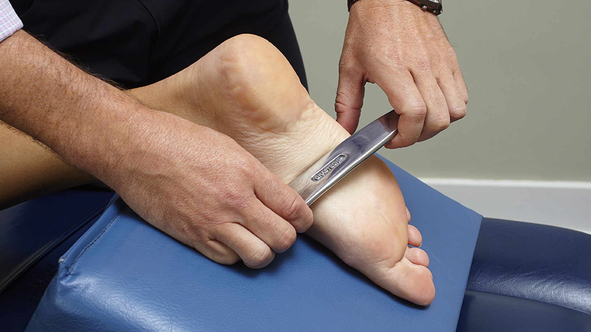
When most people think about physiotherapy, they usually think about getting treated for a muscle pain, or other strain or sprain. However, the benefits of physiotherapy extend beyond just the musculoskeletal tissues of the body. In this blog post, we discuss through some of the lesser known conditions or symptoms that can improve with physiotherapy
-
Pelvic health physiotherapy for pelvic pain or dysfunction
Firstly, pelvic health physiotherapy may be right for you if you experience pain or dysfunction in the pelvic area. This may include but is not limited to pain during intercourse, and pain or abnormal control over urination and bowel movements (urgency, incontinence, retention) and prenatal and post-natal care. Both men and women may benefit from pelvic health physiotherapy.
-
Cancer rehabilitation
Secondly, physiotherapy can help manage the many side effects of cancer treatment after surgery, radiation, and chemotherapy. Addressing these side effects can help you increase the quality of your life and get you back into your normal routine. Physiotherapy can help you with scar management, weakness, loss of range of motion, fatigue, pain, swelling, and nerve disorders after cancer treatment.
-
TMJ/jaw pain

Additionally, the temporomandibular joint (TMJ) can be a source of pain and dysfunction in individuals. Many people clench or grind their teeth without conscious thought. You may experience clicking, locking and tightness through the jaw. While most people think of the dentist when they have pain around their mouth, physiotherapists are experts in treating the TMJ. The TMJ is just like any other joint in the body. Treatment may include manual therapy, relaxation and posture exercises, correcting the jaw movements and more.
-
Vestibular physiotherapy for dizziness and vertigo

Image by cookie_studio on Freepik
Dizziness and vertigo are often symptoms that arise when there is a problem in the vestibular system of our body. Our vestibular system is in the inner ear system and is responsible for coordinating balance, head and eye movements. Vestibular rehabilitation is mostly performed by physiotherapists and a comprehensive assessment will be done to determine the correct treatment plan for your specific vestibular disorder. Treatment may include repositioning maneuvers, eye and head exercises, and exercises to retrain balance and decrease motion sensitivity
-
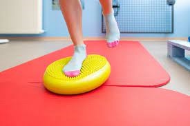
Fall prevention
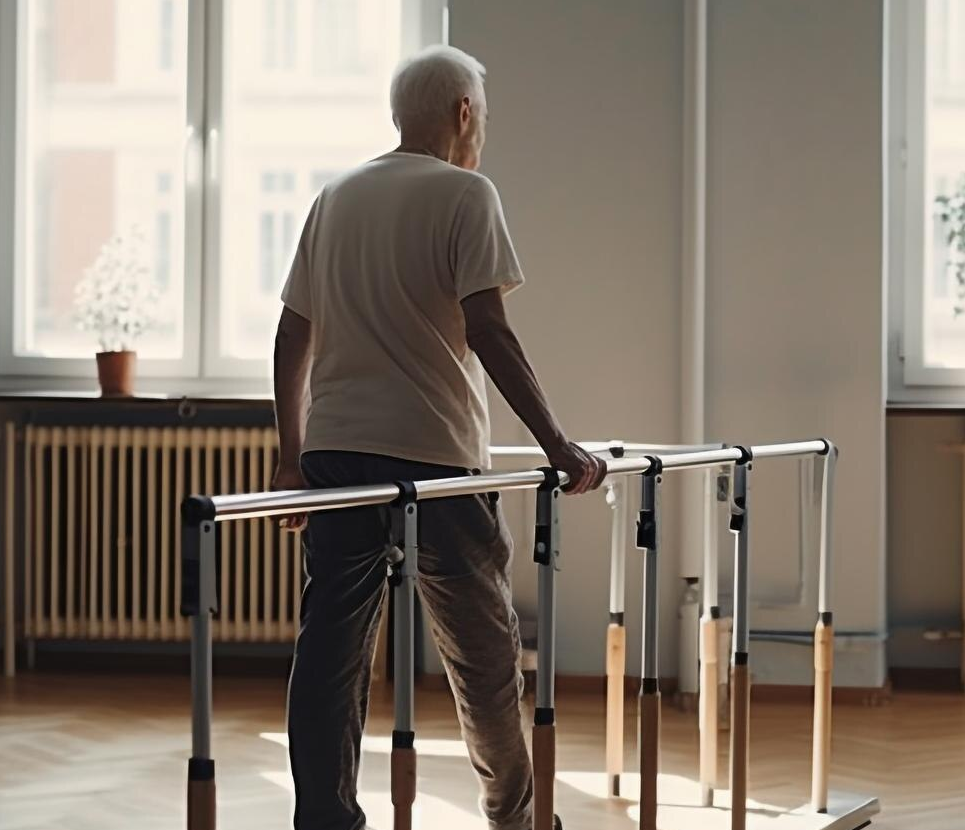
If you have had a previous fall or are concerned about your balance, then physiotherapy can help you. Your physiotherapist will work with you to determine where your risk is coming from, whether it’s dizziness, weakness, or something else. They will give you advice on how to set up your environment to minimize your risk of falling and may prescribe you a walking aid like a cane or walker. They will also give you a home exercise program to work on your balance and strengthen your muscles.
-
Osteoporosis
Osteoporosis is characterized by low bone mass, leading to an increase in bone fragility and risk of fractures. This can be scary and discourage people from their normal activities and exercises out of fear. However, with supervision from a professional like a physiotherapist, exercise therapy is one of the best treatments to minimize bone loss and restore strength. Your physiotherapist will show you how to safely perform exercises in the clinic, and give you a home exercise program to ensure that you are still able to live the life you want.
If any of these above symptoms or conditions match what you’re feeling, find us at PhysioNow. We have many expert physiotherapists who would love to help you out. Book with PhysioNow today for your first assessment and treatment!