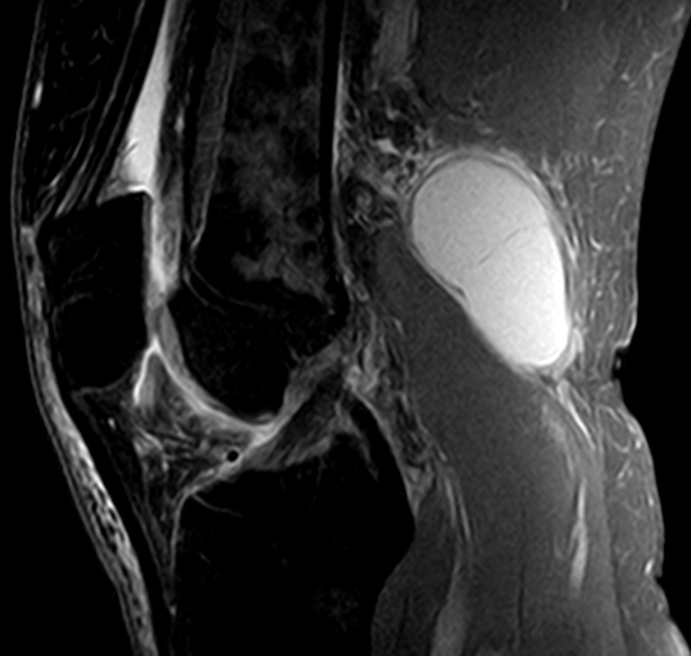
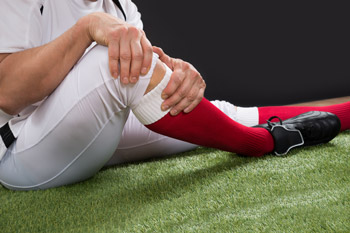
Tag Archives: Applewood Sports & Physiotherapy Ltd

ACL REHAB

ACL REHAB
Knee Injuries

ACL REHAB

ACL REHAB is very important for you to get back to your normal life! injuries lead to pain, inflammation, internal bleeding and instability.

Mechanism of Injury

The mechanism of injury is a sudden rotational force that occurs when the foot is planted on the ground.

Symptoms of ACL tear

During certain movements you may feel instability and pain such as when walking and coming down stairs.

Muscles may get weak because they respond to stress put on them during weight bearing activities.

After injuring the ACL, people tend to to avoid pain.

This can lead to decreased balance, strength and control in the muscles.

It may further lead to locking of other structures that are also involved with an ACL tear. This includes meniscus and medial collateral ligament injuries.

Is Surgical Repair required?

Surgical repair may be needed for a fully torn ACL depending on the level of physical activities you want to return to. It is possible to avoid surgery completely if proper physiotherapy is done with a skilled Registered Physiotherapist.

Custom Knee Braces for ACL REHAB
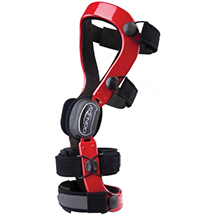
ACL REHAB
Custom Knee Brace

In addition, wearing a custom knee brace can allow you to fully support your knee without having to do surgery. Custom knee braces can be very helpful once the swelling goes down. They can allow you to return to all or most of your previous activities without surgery. A Custom Knee Brace provide the support for the torn ACL ligament. There are several types of Custom Knee braces available in the market. The most popular brands are DonJoy, Bledsoe and Ossur. At PhysioNow, we carry off the shelf knee braces and Custom Knee braces from all of the major suppliers.

Check out this link for a few of the braces we currently offer at PhysioNow in Mississauga.

How can Physiotherapy Help after an ACL tear?

Role of a physiotherapist after an ACL tear is:

– to control pain,

-increase/maintain ROM,

– stretching tight structures,

and strengthen the weak muscles

How is Pain controlled?

Pain is controlled by using different modalities such as ultrasound and interferential therapy. A graduated exercise program will help you to improve your movement.
Here is another interesting article about ACL REHAB.

Exercises will focus mainly on:

– strengthening quadriceps and gluteal muscles,

– proprioception techniques (you will likely lose some of your natural ability to balance after an ACL tear),

-and stretching of the hamstring muscles.

ACL REHAB
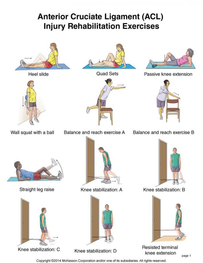
Possible ACL REHAB Exercises

Please do not attempt these exercises at home without the advice of your doctor or a skilled Registered Physiotherapist!

If you or someone you know has had an ACL tear, we at PhysioNow would be happy to help! If you want to discuss Physiotherapy Treatment or to see if a Custom Knee brace might be helpful for you, please call and ask to speak with Sharon. Call today!

PELVIC FLOOR: POSTURE

PELVIC FLOOR: POSTURE

Pelvic Floor: Posture

PELVIC FLOOR: POSTURE is a very important area if you have issues with urinary incontinence, pelvic pain, etc.  The muscles of the pelvic floor support the abdominal and pelvic viscera.  These muscles are active in standing and sitting. Furthermore, because the abdomen is a fluid-filled cavity, intra-abdominal pressure  is distributed in all directions.  The Pelvic Floor muscles, which form the floor of the abdominal cavity, contribute to its muscle control.

As a result of this contribution to control of intra-abdominal pressure, the muscles of the pelvic floor are likely to contribute to control of the spine and pelvis. The slouched posture places a lot of pressure on our internal organs and pelvic floor. This can cause a worsening of:

  • urinary incontinence,
  • pelvic pain,
  •  symptoms related to prolapse,
  • and rectus diastasis.

Pelvic Floor: Posture, sitting slumped

  • Research has shown that when we sit in a slumped posture, our pelvic floor muscle activity is much less than when we are sitting tall.
  • Pelvic Floor: Posture IS VERY IMPORTANT!
  • slouched sitting postures decrease the activity of your transverse abdominal muscles.
  • A Proper breathing pattern encourages the pelvic floor to move more dynamically.
  • Dysfunctional breathing patterns inhibit this dynamic movement of the pelvic floor.

Pelvic Floor: Posture,  

  • An assessment by a Pelvic Health Physiotherapist includes a thorough postural examination.
  • Your Pelvic Health Physiotherapist will be able to identify your unique postural compensations
  • They can help guide you on the road to improved body posture awareness.

CHECK OUT THIS LINK FOR FURTHER INFORMATION.

At PhysioNow, we have fully trained Pelvic Health Physiotherapists that can help to assess and treat pelvic issues like the following conditions:

  • Constipation
  • Dyspareunia
  • Endometriosis
  • leakage of urine
  • pain with intercourse
  • pelvic organ prolapse
  • pelvic pain
  • urinary incontinence
  • vaginismus
  • erectile dysfunction

If you or someone you know suffers from one of these conditions, give us a call today.  We would be happy to help!

  1. Sapsford, RR. et al (2006) Sitting posture affects pelvic floor muscle activity in parous women: an observation study. Aust L Physiother. 52(3):219-22
  2. Reeve, A., Dilley, A., (2009) Effects of posture on the thickness of Transverse Abdominal Muscle and Pelvic Floor Muscle Exercises for Stress Urinary Incontinence: A Randomized controlled Trial. J Phys Ther Sci. 26(8): 1161-1163.

Neck Pain Treatment

Neck Pain Treatment

Neck Pain Treatment

Pain in your neck can happen for several different reason  . Neck Pain Treatment can help get you back on your feet quickly.  It can happen suddenly from an accident or fall.  Even sleeping in an awkward position can cause neck pain. Other times it can build up over several months or even years and gradually get worse. And sometimes, it may seem like there is no reason and you just wake up one day with neck pain.

Neck Pain Treatment:  Possible symptoms you may feel

  • The way you feel the pain can also vary depending on how the  injury happened.
  • Some neck injuries will have just pain as the symptom.
  • Other neck injuries will include pain but also have weakness and pins and needles as well.
  • Sometimes you will have pain that is just in the neck itself. In other cases, pain is felt more in the arms or hands.  When this happens,  you might not think that there is anything wrong with your neck at all.
  • Neck pain Treatment can be difficult mostly because it can be difficult to properly diagnose. It is important to find an experienced Registered physiotherapist to make sure that appropriate treatment is received.

See this link for possible causes of Neck Pain.

Neck pain Treatment options:

There can be a variety of treatment options that can be successful at managing neck pain.

  • Modalities such as heat and IFC/TENS can help with reducing pain temporarily.
  • Manual treatment techniques like soft tissue work and mobilizations help with improving mobility and reducing stiffness and tissue tension.
  • Doing the appropriate exercises will also help stretch and strengthen the affected areas.
  • Acupuncture is another modality that can help with reducing pain and improving stiffness.
  • Massage therapy is another treatment approach that can greatly alleviate pain and stiffness and help further the patient’s recovery.
  • Ultimately, there are a variety of treatment options available to treat neck pain. It is up the treating therapist and their expertise to determine the best treatment approach to correct the pain and achieve the patient’s goals.

Neck pain can be very painful  and really affect a your quality of life. It is important to seek help as soon as possible so that the pain and limitations can be addressed and recovery can be achieved quickly!

Call PhysioNow today to get started!

Baker’s Cyst

Baker's cyst

A Baker’s Cyst, also known as a popliteal cyst, is a fluid-filled swelling that causes a lump at the back of the knee. This leads to tightness and restricted movement. The Baker’s Cyst can be painful when you bend or straighten your knee.

? Causes of a Baker’s Cyst

Synovial fluid is a clear liquid that normally circulates through the spaces in your knee joint. Sometimes the knee produces too much of this fluid. The increasing pressure forces the fluid to the back of the knee via a one-way valve. This creates a bulge. This  swelling of the knee causes a Baker’s Cyst to form.

The most common causes of a Baker’s Cyst are:

  • damage to the knee’s cartilage (meniscus)
  • arthritis of the knee
  • rheumatoid arthritis
  • other knee conditions that cause joint inflammation

? Symptoms of a Baker’s Cyst

Some patients may have no pain and may not even notice the cyst is there. However, the signs and symptoms of a Baker’s cyst can include:

  • Swelling or lump behind the knee (the main sign) – this is more evident when standing and comparing one knee to the other. It may feel like a water-filled balloon.
  • Knee pain.
  • Calf pain.
  • Accumulation of fluid around the knee.
  • The knee joint may click or buckle.
  • The knee joint may lock.
  • ? Diagnosis of a Baker’s Cyst

    • Your doctor will examine your knee and feel the swelling. If the cyst is small, they may compare the affected knee to the healthy one and check your range of motion.
    • Your doctor may recommend non-invasive imaging tests if the cyst rapidly increases in size or causes severe pain or fever. These tests include an MRI or ultrasound. An MRI will enable your doctor to see the cyst clearly and to determine if you have any damage to the cartilage.
    • These tests will determine if some other form of growth, such as a tumor, is causing the swelling.
    • Although the cyst won’t show up on an X-ray, your doctor may use one to check for other problems, such as inflammation or arthritis.

    Web MD has additional info on this condition.

    ? Treatment

    A Baker’s Cyst doesn’t need treatment. It will go away on its own. However, if the swelling becomes large and causes severe pain, your doctor may recommend one of the following treatments.

    • Fluid draining

      Your doctor will insert a needle into the knee joint and may use an ultrasound to help guide the needle to the correct place. They’ll then draw the fluid from the joint.

    • Medication

      Your doctor may recommend a corticosteroid medication, such as cortisone. Your doctor will inject this drug into the joint, and the medication will flow back into the cyst. Though it may help relieve the pain, it doesn’t always prevent a Baker’s Cyst from recurring.

    • Physiotherapy

      Regular, gentle exercises may help increase your range of motion and strengthen the muscles around your knee.

    If you think you or someone you know may have a Baker’s Cyst, please call PhysioNow today!  We would be happy to help! We have highly trained Registered Physiotherapists ready to see you Now!

Acupuncture: Neck pain

Acupuncture: Neck pain

Acupuncture: Neck pain

Almost everyone will experience some sort of neck pain or stiffness during their lifetime. However, certain occupations appear to be predisposed to neck symptoms.  Acupuncture: Neck pain may be able to help.

Manual laborers, for instance, have more symptoms than office workers.  The type of work seems to affect the risk. A person’s age and a history of twisting and bending during work can also contribute.

Studies have shown acupuncture: Neck Pain to be effective in relieving certain types of neck pain.  This is especially true for neck pain  caused by whiplash. Some studies suggest acupuncture can treat degenerative neck disorders such as ankylosing spondylosis and cervical spondylosis.  In many cases, acupuncture: Neck pain has worked for patients whose conditions could not be solved using conventional approaches.

¬ Mechanism of action of Acupuncture: Neck pain

  • Release of opioid peptides.Opioids are naturally occurring chemicals in the brain that have apain killing effect. The release of these opioids plays a large role in the reduction of pain. There has been lots of evidence to show that acupuncture stimulates the central nervous system.  This causes the release of these pain killing chemicals.
  • Alteration in the secretion of neurotransmitters and neurohormones.Acupuncture is said to activate the hypothalamus and pituitary glands. This changes the release of these chemicals. These particular chemicals play a direct role in the feeling of pain. It also affects the activity of an organ . Evidence has shown that acupuncture alters this secretion in a manner that reduces pain.
  • Stimulation of electromagnetic points on the body. The 2,000 points of the body that acupuncture focuses on are thought to be special conductors of electromagnetic signals. Stimulation of these areas is believed to start the flow of endorphins—the body’s natural painkillers.¬ Indications for Acupuncture: Neck pain
    • Sudden Force/Automobile Accident
    • Degenerative Disc Disease
    • Overuse/Improper Use
    • Osteoporosis
    • Neck Strain
    • Degenerative Arthritis
    • Whiplash
    • Muscle Tension or Spasm
    • Bone Spur
    • Herniated or Protruding Disk
    • Pinched Nerve
    • Ligament/Muscle Tears
    • Cervical Spondylosis
    • Ankylosing Spondylosis

    ¬ Contraindications of Acupuncture: Neck pain

    There are very few situations where acupuncture is not advised:

    • When someone has a hemophilic condition
    • When a  patient is pregnant – certain acupuncture points and needle manipulations should not be used during pregnancy
    • If a patient has a severe psychotic condition or is under the influence of drugs or alcohol

    Check out this link for a bit more information about the use of Acupuncture: Neck pain.

    ¬ Types of application of Acupuncture: Neck pain

    • Basic needlingNeedles are inserted to a depth of 4–25 mm and left in place for a period of time (from a few seconds to many minutes). There are often 6–12 needles (and sometimes more) inserted at different acupoints at the same time. The sensation is often described as a tingling or dull ache at the entry point. Many people say they feel very relaxed or sleepy, and some report increased energy levels afterwards.
    • Electro acupuncture (EA)A tiny focused electric current is applied to the skin at the acupoints or can be applied to the needle itself.

    If you would like to meet with a Registered Physiotherapist that is trained to do Acupuncture: Neck Pain,   please call PhysioNow today! Our experienced physiotherapists would be happy to help !

Peroneal tendinopathy

Peroneal tendinopathy

Peroneal tendinopathy

Peroneal tendinopathy or peroneal tendonitis is characterized by an aching pain and swelling in the perineal tendons. These are located in the lower, outside portion of the ankle. A tendon is soft-tissue that attaches a muscle to a bone. The muscles involved in this condition are the 2 peroneal muscles in the lower leg, called the peroneus longus and the peroneus brevis.

Anatomy

?There are two peroneal tendons that run along the back of the fibula. The first is called the peroneus brevis. The term “brevis” implies short.  It is called this because it has a shorter muscle and starts lower in the leg. It then runs down around the back of the bone called the fibula on the outside of the leg and connects to the side of the foot.  The peroneus longus takes its name because it has a longer course. It starts higher on the leg and runs all the way underneath the foot to connect on the other side of the foot. Both tendons, however, share the major job of turning the ankle to the outside. The tendons are held in a groove behind the back of the fibula bone.

Causes of Peroneal Tendonitis

  • A sudden increase in weight bearing activities, particularly walking, running or jumping
  • Inadequate or unsupportive footwear
  • Muscle imbalances of the lower limb
    Poor lower limb biomechanics
  • Incomplete rehabilitation following an acute ankle injury, such as an ankle sprain

Symptoms of Peroneal Tendinopathy

  • Gradual worsening pain over the outside of the ankle
  • Pain during and/or after weight bearing activities
    Pain with turning the foot in and/or out
  • Instability around the ankle when weight bearing

Diagnosis

A full examination from a physiotherapist can be all thats needed to diagnose peroneal tendonitis
Patients with this condition usually experience pain behind the outside ankle during activities putting stress on the perineal tendons. Pain can also be noticed following these activities or following a rest period. This may be noticed especially upon waking in the morning. There may be swelling when the injury first happens. There will also be pain when testing resisted foot movements.  Stretches into various positions of the foot inversion, and resisted movements can cause pain behind the outside ankle.
Diagnosis may be confirmed with an MRI scan or ultrasound investigation
a diagnostic Ultrasound may be used for detecting all types of peroneal injuries.

What else could it be?:

Symptoms of peroneal tendinopathy mimic various other conditions of the ankle joint. So, before diagnosing peroneal tendinopathy we should rule out other possible injuries by doing the following tests:
Ankle Sprain: ligament testing by the Physiotherapist
Ankle fractures: special tests by the Physiotherapist
Os trigonum syndrome: MRI, physiotherapy testing
Chronical lateral ankle pain with other cause: MRI
Longitudinal peroneal tendon tear: MRI
Peroneal subluxation: ultrasonography, CT, MRI or peroneal tenography
Flexor Hallucis longus tendon injury

Physiotherapy rehabilitation

Treatment for peroneal tendonitis includes a program of stretching, strengthening, mobilisation and manipulation. It also includes proprioceptive exercises, icing, ankle bracing or k-taping during contact sports. If symptoms are severe, a cast or ROM boot immobilization may be worn for 10-20 days. After symptoms resolve, you will begin a progressive rehabilitation programme along with a gradual increase to full activity.

The use of a biomechanical ankle platform (BAPS), deep tissue friction massage, ultrasound electric stimulation can also be included in the physiotherapy
Also, shock wave therapy (ESWT), acupuncture is used to treat tendinopathy. But there is only limited evidence from studies for these treatments.
There is evidence for using manual therapy, specifically the lateral calcaneal glide.

If you have any further queries please call PhysioNow. Our experienced physiotherapists would be happy to help you. Call Today to get started 289-724-0448.!

De Quervain’s Disease

De Quervain’s Disease/ Texting thumb.

De Quervain's Disease

De Quervain’s Disease

De Quervain’s Disease or nowadays known as texting thumb is a painful inflammation of tendons in the thumb that extend to the wrist. The rubbing of the inflamed tendon against the canal it passes through causes pain at the base of the thumb and into the lower arm. It is commonly seen in females over 40 years of age.

Causes of De Quervain’s Disease

1. Simple strain injury to the tendon.
2. Repetitive motion injury. Workers who perform rapid repetitive activities involving pinching, grasping, pulling or pushing are at increased risk. Specific activities including intensive mousing, trackball use, and typing. Other activities including bowling, golf, fly-fishing, piano-playing, sewing, and knitting can also cause De Quervain’s Disease.
3. Frequent causes of De Quervain’s Disease include stresses such as lifting young children into car seats, lifting heavy grocery bags by the loops, and lifting gardening pots .
4. De Quervain’s Disease often occurs during and after pregnancy. Factors may include hormonal changes, fluid retention and more lifting.
5. Rheumatoid arthritis.

De Quervain's Disease

De Quervain’s Disease/ Texting Thumb

Onset and Symptoms of De Quervain’s Disease

Onset can be gradual or sudden. Pain is felt along the back of the thumb. There can be Pain directly over the thumb tendons, and pain may travel into the thumb or up the forearm. The bottom of the thumb or the side of the wrist might also be sore or swollen.
It may be hard and painful. Symptoms may get worse when the thumb is moved, particularly when pinching or grasping things. Some people also have swelling and pain on the side of the wrist at the base of the thumb. The back of the thumb and index finger may also feel numb. People might experience a funny sound like a squeak, crackle, snap, or creak when they move the wrist or thumb.
If the condition is not well addressed, the pain can spread up your forearm or down into your thumb.

How is De Quervain’s Disease diagnosed?

De Quervain’s Disease is diagnosed based on history and physical examination. X-rays, or ultrasound may be used to rule out other causes of pain.The Physiotherapist may use special tests to help diagnose De Quervain’s. More information can be found here.

Physiotherapy Treatment

Your Physiotherapist will likely recommend that you wear a specific wrist splint with a thumb spica for 4 to 6 weeks . PhysioNow carries these wrist splints in stock. You’ll also need to stop doing activities that worsen the condition.

Wrist brace with thumb spica

De Quervain’s Disease

The physiotherapist after a thorough assessment could choose different therapy approaches to help with the swelling, pain, and function.
These treatments may include Ultrasound, K-taping, acupuncture, and manual therapy. Other Physiotherapy treatments may include specific exercises focusing on range of motion, strength, and flexibility. These would be given for a safe and effective return back to function.

Recovery times vary depending on your age, general health, and how long you’ve had the symptoms.
If your disease has developed gradually, it’s often tougher to treat. So, it may take you longer to get relief. Your doctor may give you anti-inflammatory medication, or may inject the area with steroids to curb pain and swelling.

In our experience at PhysioNow, more than 99% of people with De Quervain’s Disease get better with Physiotherapy treatment provided. If however, you are one of the outliers, your doctor may recommend surgery. The operation would release the tendon’s tight covering so that the tendon could move smoothly. It’s an outpatient procedure, which means you go home afterward. Your doctor will recommend physiotherapy after surgery which includes an exercise program to strengthen your thumb and wrist.

If you or someone you know suffers from De Quervain’s Disease, please call us today. Our skilled Physiotherapists can Help!

Vaginismus

Vaginismus

Vaginismus and Physiotherapy

Vaginismus is a painful feeling of discomfort or inability when inserting a tampon, finger, penis or during a doctor’s internal pelvic exam. It occurs when there are involuntary contractions of the muscles in the outer third of the vagina.

Primary Vaginismus: when a woman has never been able to have pain free intercourse due to pelvic floor muscle spasm.

Secondary Vaginismus: pain that develops sometimes later in life after a traumatic event such as childbirth, surgery, or a medical condition.

With Vaginismus, there is usually significant Connective Tissue Dysfunction that needs to be addressed first before any internal work. It is suggested that you follow up the self-help treatment for connective tissue dysfunction before embarking on the stretching exercises with the dilators.

Pelvic floor exercises and Desensitisation techniques

A physiotherapist may be able to teach you pelvic floor exercises, such as squeezing and releasing your pelvic floor muscles, that can help you gain control over the muscles causing the vagina to close involuntarily. Occasionally, a technique called biofeedback may be useful. A small probe is inserted into your vagina. This monitors how well you are doing the exercises by giving you feedback as you do them.
Biofeedback can help with the following:

• Teach you strategies that may help to relax the muscles that are contracting involuntarily
• Teach you appropriate timing of the contraction and relaxation of the pelvic floor muscles.
• Educate you about your condition and why it might be occurring.
• Teach you various strategies for getting the muscles to relax
• help you to Use manual therapy to release the muscles either externally and/or internally that are causing the contractions.

More information about Vaginismus can be found here.

Some general tips about pelvic floor muscle exercises:

• Choose a convenient time and place where you can exercise regularly.
• Put a reminder about pelvic muscle exercises somewhere obvious to you (or set an alarm on your watch or mobile phone).
• Avoid quick pelvic floor muscle exercises where you don’t hold the contraction. This tends to cause an increase in pelvic floor muscle resting tone.
• Be aware that it will take 6 to 8 weeks of regular exercise before you can expect to see improvement. It will take approximately 3 months for the pelvic muscles to strengthen.
• You could try inserting one finger into the vagina to check the strength of your squeeze as you pull in the pelvic muscles. Alternatively, you may be able to feel a pelvic floor contraction if you place the tip of your index finger on the perineal body (on the outside, between the vagina and back passage).

Other tips to reduce Vaginismus:

• If you are overweight, try reducing your weight.
• Try not to become constipated, as a full bowel will put pressure on the bladder. Straining to empty your bowel will weaken the pelvic muscles.
• If your job involves lifting, think of your pelvic muscles as well as your back. Pull up your pelvic muscles when lifting a heavy weight.
• If you smoke, consider quitting. Frequent coughing puts a strain on the pelvic muscles.

If you or someone you know suffers from Vaginismus, please call PhysioNow today! Our specially trained Physiotherapist can start your treatment Now!

Dyspareunia

Dyspareunia and Physiotherapy
Dyspareunia
Dyspareunia is genital pain experienced by women just before, during or after sexual intercourse. Some women have always experienced pain with intercourse from their very first attempt. Other women begin to feel pain with intercourse or cyclically with menstruation. They can also have pain after an injury or infection . Sometimes the pain increases over time. When pain occurs, the woman may be distracted from feeling pleasure and excitement.

Causes

• vaginal dryness from menopause, childbirth, breastfeeding, medications
• skin disorders that cause ulcers, cracks, itching, or burning
• infections, such as yeast or urinary tract infections
• spontaneous tightening of the muscles of the vaginal wall
• endometriosis
• pelvic inflammatory disease
• uterine fibroids
• irritable bowel syndrome
• radiation and chemotherapy

Other factors that affect a woman’s ability to become aroused can also cause dyspareunia.
These factors include:
• stress, which can result in tightened muscles of the pelvic floor
• fear, guilt, or shame related to sex
• self-image or body issues
• medications such as birth control pills
• relationship problems
• cancer, arthritis, diabetes, and thyroid disease
• history of sexual abuse

Symptoms of Dyspareunia

• Pain while inserting a tampon or during penis penetration
• Pain with certain sexual partners
• Deep pain during thrusting
• Burning pain or aching pain
• Throbbing pain, lasting hours after intercourse

Check out this link for more information.

Physiotherapy Treatment for Dyspareunia

Education:

To help describe how the pelvic floor muscles can cause pain. Education can also provide techniques that can be used at home.

Manual Physiotherapy: to mobilise muscle and soft tissue, normalize overactive muscles, improve circulation and desensitize painful areas.

Desensitization therapy: learning vaginal relaxation techniques, such as Kegel exercises, that can decrease pain.

Sex therapy: learning how to re-establish intimacy and improve communication with your partner.

Water-based lubricants rather than petroleum jelly or other oil-based lubricants are preferable. Oil-based lubricants tend to dry the vagina.

Psychologic therapies: Cognitive-behavioral therapy and mindfulness-based cognitive therapy can be helpful.

Pelvic muscle relaxation exercises: used with biofeedback, this can help women with tight pelvic muscles learn to consciously relax tight muscles.

If you or someone you know suffers from dyspareunia, please call today to get started on Physiotherapy treatment! We can help at PhysioNow. Often only a few visits with our specially trained Phyiotherapist can help to get you on the road to recovery.

Hip fracture

Anatomy of Hip
A hip fracture is a break in the upper quarter of the femur (thigh) bone. The extent of the break depends on the forces that are involved. The type of surgery used to treat a hip fracture is based on the bones and soft tissues affected or on the level of the fracture.
Older people are at a higher risk of hip fracture because bones tend to weaken with age (osteoporosis). Multiple medications, poor vision and balance problems also make older people more likely to trip and fall — one of the most common causes of hip fracture.

? Signs and symptoms of a hip fracture include:
• Inability to move immediately after a fall
• Severe pain in your hip or groin
• Inability to put weight on your leg on the side of your injured hip
• Stiffness, bruising and swelling in and around your hip area
• Shorter leg on the side of your injured hip
• Turning outward of your leg on the side of your injured hip

? Causes of Hip fracture
• falling on a hard surface or from a great height.
• blunt trauma to the hip, such as from a car crash.
• diseases such as osteoporosis, which is a condition that causes a loss of bone tissue.
• obesity, which leads to too much pressure on the hip bones.

? Types of Fractures
In general, there are three different types of hip fractures. The type of fracture depends on what area of the upper femur is involved.
Intracapsular Fracture
These fractures occur at the level of the neck and the head of the femur, and are generally within the capsule. The capsule is the soft-tissue envelope that contains the lubricating and nourishing fluid of the hip joint itself.

Intertrochanteric Fracture

This fracture occurs between the neck of the femur and a lower bony prominence called the lesser trochanter. The lesser trochanter is an attachment point for one of the major muscles of the hip. Intertrochanteric fractures generally cross in the area between the lesser trochanter and the greater trochanter. The greater trochanter is the bump you can feel under the skin on the outside of the hip. It acts as another muscle attachment point.

Subtrochanteric Fracture

This fracture occurs below the lesser trochanter, in a region that is between the lesser trochanter and an area approximately 2 1/2 inches below .

In more complicated cases, the amount of breakage of the bone can involve more than one of these zones. This is taken into consideration when surgical repair is considered.

Treatment for hip fracture: usually involves a combination of surgery, rehabilitation and medication.

? Surgery
The type of surgery you have generally depends on the location and severity of the fracture. Are the broken bones properly aligned? (displaced fracture. What is your age? What are your underlying health conditions?

The options include:

• Internal repair using screws. Metal screws are inserted into the bone to hold it together while the fracture heals. Sometimes screws are attached to a metal plate that runs down the upper thigh.

• Partial hip replacement. If the ends of the broken bone are not lined up or damaged, your surgeon may remove the head and neck of the femur and install a metal replacement (prosthesis).

• Total hip replacement. Your upper thigh and your hip socket are replaced with an artificial one (prostheses). Total hip replacement may be a good option if arthritis or a prior injury has damaged your joint. This may have been affecting your hip function even before the fracture.

? Rehabilitation

Rehabilitation is begun as soon as possible after hip fracture surgery, often within a day. The initial goals are to help people retain the level of strength they had before the fracture. You want to keep moving to prevent loss of muscle. You also want to prevent problems that result from bed rest. The ultimate goal is to restore your ability to walk properly without a limp.

Benefits of Rehab

Rehab will help you:

• Restore normal movement in your joint
• Build up strength in the joint and surrounding muscles
• Ease pain and swelling
• Let you get back to your normal activities including walking without a limp
• Help with circulation, particularly right after surgery, so you don’t have problems with blood clots

If you have or someone you know has fractured a hip or had hip replacement surgery, please call PhysioNow. Our experienced physiotherapists would be happy to help with your recovery! Call today to book an appointment!