Tag Archives: Foot Pain Management Mississauga

Few things to know about your heel pain

What is heel pain?

Heel pain

What causes your heel pain?

Heel pain could be from any reason either your inflamed plantar fascia causing plantar fasciitis or any abnormal bony growth causing heel spurs. To get in further details plantar fasciitis is an irritation and inflammation to the tissue that runs at the bottom of the foot extending from the heel to the toes, causing heel pain. It supports the arch of the foot which helps in shock absorption. On other side, heel spur is when a bony outgrowth forms on the heel, which causes heel pain while walking and standing.

What are the causes of heel pain?

Causes are not limited to high and low arches causing irritation to the plantar fascia with running, jogging, playing any sports, prolonged standing and walking. Also, any strain or tear to the fascia. As a result, if this condition is left untreated can lead to bony growth causing heel spurs.

5 Most common risk factors of pain

  • Improper foot wear: The foot wear without proper arch support or worn-out shoes.

    Causes of heel pain

    Improper footwears

  • Weight gain leads to changes in arches and support and affects the weight transmission and shock absorption.
  • Foot mechanics: flat feet or high arches, stiff mid foot and ankle.

    Risk factors for plantar faciitis

    Types of Arches

  • Age: commonly seen between 40 – 60 years old.
  • Occupation: commonly seen in clients with jobs that requires long standing.

What are the symptoms:

With plantar fasciitis it is like a stabbing pain felt at arch of foot and near the inner part of heel whereas pain with spurs is in the center of heel and also feels like a stabbing.

Heel spurs and plantar fascia

Location of pain

Pain is usually worse in the morning taking first few steps with plantar fasciitis. It usually gets better with walking but comes back with long hours of standing and sitting. On other hand pain with spurs gets worse as the day progresses with long hours of standing and walking. Sometimes, there could be swelling present over the heel area.

How will Physiotherapy help you with heel pain?

Your physiotherapist after a detail assessment would be able to identify the cause of the problem and give you advice on how to modify and handle the situation and life style which will help in the road to recovery. Advice would include,

Arch supports

Foot insoles for arch support

  • How to modify activities of daily living.
  • Advice on foot wear.
  • Suggestions on arch supports and custom-made orthotics, night splint.

Apart from this, treatment sessions will include:

  • Hands on techniques to release the tight plantar fascia, calf, mobilization of foot and ankle manually or with a tool.  Stretches for the tight surrounding structures.

    Techniques to release plantar fascia

    Hawk tool to release plantar fascia

  • Strengthening exercise for the supporting structures and muscles that maintain the arches and functional training
  • Kinesiotapping to relieve stress from the affected structures.

Don’t let your pain affect your quality of life and stop you from doing your activities. Book your assessment session now with qualified physiotherapists of PhysioNow. We have various locations to serve the communities in Mississauga, Oakville and Etobicoke. Contact us and get started today!

Acute Ankle Sprains

Acute Ankle Sprains

Acute Ankle Sprains

Acute Ankle Sprains

How do Acute Ankle Sprains happen?

Acute Ankle sprains are common injuries in people of all ages. They can happen in many different circumstances. However, they always involve some type of fall or twisting movement to the ankle. For example:

  • Going downstairs and missing the last step resulting in twisting the ankle
  • Landing on the ankle in a twisted position after coming down from a lay-up in basketball or in a Sports Injury
  • Slipping on an icy street while walking

In all of these situations, the ankle gets a sudden movement and/or trauma that causes it to twist more than its normal range of movement allows.

Acute Ankle Sprains

Acute Ankle Sprains

What structure gets injured with an acute ankle sprain?

The structures that are injured with acute ankle sprains are the ligaments around the ankle joint. Ligaments are soft tissue structures that connect two bones together. Their function is to prevent the bones from moving too far away from each other. Ligaments help maintain normal movement between the bones.

With ankle sprains, the ligaments get over-stretched beyond what they can tolerate which causes tears in their fibres. The ligaments that are involved in acute ankle sprains are present on the outside (lateral), inside (medial), and top of the ankle.

Are there different types of acute ankle sprains?

There are three main types of ankle sprains:

  • Inversion
  • Eversion
  • High Ankle Sprain

The terms relate to the direction of ankle movement that caused the injury to occur.

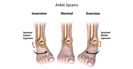
INVERSION ANKLE SPRAIN

  • An inversion sprain occurs when the ankle is forcefully turned inwards.
    • This type of acute ankle sprain is the most commonly occurring of the three because the ligaments on the outside of the ankle are generally weaker than the rest.
    • Another name for an inversion ankle sprain is lateral ankle sprain
    • A lateral ankle sprain injures the ligaments on the outside of the ankle.

EVERSION ANKLE SPRAIN

  • An eversion sprain occurs when the ankle is forcefully turned outwards.
    • This type of force injures the ligaments on the inside of the ankle.
    • The ligaments on the inside of the ankle are called the deltoid ligament
    • An eversion ankle sprain is also called a medial ankle sprain

HIGH ANKLE SPRAIN

  • A high ankle sprain is the rarest of the three types and it occurs when there is excessive twisting of the shin.
    • The ligaments that are injured with this type of injury are located above the ankle.
    • The location of the ligaments is why this injury is called a ‘high’ ankle sprain.


Are some people more at risk for Acute Ankle Sprains than others?

There are certain risk factors that may make ankle sprains more likely to happen. However, the research is a little inconclusive on why this is so. Some of these possible factors include:

  • Poor muscle strength/fitness
  • Increased weight
  • Decreased muscle-reaction time
  • Previous history of ankle sprains resulting in poor balance

Can there be different severities of Acute Ankle Sprains?

The severity of the ankle sprain depends on how much the ligament was overstretched in the process of the injury. Sprains are classified by different grades to explain the severity:

  • Grade I: mild stretching or tear of the ligament, with little or no instability at the joint
  • Grade II: incomplete tear of the ligament, with some instability in the joint
  • A Grade III: ligament is completely torn or ruptured and there is no stability in the ankle
Acute Ankle Sprains

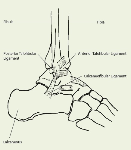
Ligaments affected in Ankle Sprains

What should you do if you think you have sprained your ankle?

In all three types of Acute Ankle Sprains, it is important that you see a health care provider. The health care provider, whether it is a doctor or a Registered Physiotherapist, will first rule out the possibility of fracture. There is always the possibility of a fracture with Acute Ankle Sprains. The ligaments attach onto a bony area and if the twist is severe enough, it can pull off a chunk of the bone.  When this happens, it can cause a fracture. Signs and symptoms of a potential fracture include:

  • Lots of swelling
  • Increased bruising
  • Increased tenderness over specific areas of the ankle
  • Difficulty or inability to put weight through the injured ankle

When the signs and symptoms are suspicious of a fracture, then an X-ray must be completed. If there is no evidence of fracture, then your doctor may send you for an ultrasound.  In the end,  they should refer you for Physiotherapy treatment.  So ask your doctor to send you to Physiotherapy if they don’t think to recommend it themselves.

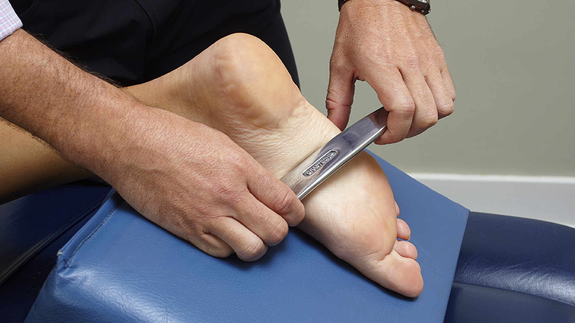
What will happen during my first Physiotherapy appointment?

During the Physiotherapy assessment, your Registered Physiotherapist will assess everything to do with your Acute Ankle injury, including:

  • Pain intensity and location
  • Swelling
  • Sensation
  • Range of motion
  • Ankle stability
  • Strength
  • Balance

This will provide some baseline information for your Physiotherapist.

How can Registered Physiotherapy help treat Acute Ankle Sprains?

The treatment for acute ankle sprains depends on the type of sprain as well as the severity of the injury.

Immobilization

  • Because of their affect on ankle stability, a Grade II or III injury will require some immobilization. This is usually done through a removable walking cast.
  • The purpose of the immobilization is to protect your injured ligaments from movement and external stressors, so that they can heal.
  • The period of immobilization will depend on the injury and your health care provider will provide specific instructions.
  • A grade I sprain will not require any immobilization because the stability of the ankle is still intact. However, your Physiotherapist will recommend activity modification to help with the healing process.

Physiotherapy Treatment in the acute phase?

  • In the initial phase of the treatment, the Physiotherapist may focus on pain relieving modalities
  • Electrical modalities like IFC and Ultrasound can help relieve pain and swelling
  • The Physiotherapist will also prescribe gentle mobility exercises to help improve or maintain movement through the ankle
  • The length of the acute phase depends on the severity of the sprain
Registered Physiotherapy for Acute Ankle Injuries

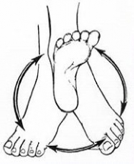
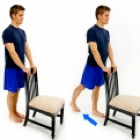
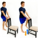
Ankle Rotations for movement Recovery

Treatment in the later stages of Acute Ankle Sprains

  • Once the ligament healing is underway and the patient feels a little better with their pain, the treatment will get more progressive
  • It will focus on regaining mobility, strength and function through the ankle
  • Your Physiotherapist will prescribe exercises that are going to help regain the lost range of motion and strength.
  • Over several weeks, the exercises will be progressed as you improve.
  • Another important aspect of treating an ankle sprain is including balance re-training in the program.
  • Balance is very much affected after an ankle sprain and it is something that must be improved with specific exercises. This is very important no matter whether you are returning to Sports or just everyday living!  Balance Retraining is key to Falls Prevention!
  • We use our balance ability on a regular basis with walking on uneven surfaces or when trying to recover from a stumble. Therefore, it is important to improve balance ability to prevent any possible re-injury of the ankle.
  • If you have had an acute ankle sprain before, and DID NOT attend Physiotherapy, it is likely that you are at increased risk of reinjuring your ankle or falling again!
Acute Ankle Injuries

Physiotherapy Mobilizations

Do I need to get Physiotherapy treatment after Acute Ankle Sprains?

There are some people who have sprained their ankle multiple times and never sought out treatment for the injury. They usually give themselves some time for the pain to get better and then go about their usual activities.

This type of management is not usually recommended because multiple ankle sprains can affect the stability of your ankle. As a result, this may create problems in the future. Also, the reduced stability in your ankle may cause compensations in your knees and hips. This can complicate the picture and make your injury more complex.

It is therefore recommended that you seek out proper Registered Physiotherapy care to address an ankle sprain soon after it occurs. This ensures that your acute ankle sprain heals in a timely manner without added complications.  The most important part of treatment is regaining your balance.  If you do not do this, another injury in quite likely.

To get started on your Acute Ankle Sprain Treatment, Give PhysioNow a call Today!

Patellofemoral Pain Syndrome

Patellofemoral pain syndrome

Patellofemoral Pain Syndrome

Patellofemoral pain syndrome happens when your knee cap does not track properly.  The knee joint consist of two joints.  The tibiofemoral joint which is between the two long bones in your leg and the second one is the patellofemoral joint.  This  is between the femoral condyle (end of thigh bone) and the knee cap. The cartilage on the back of the knee cap glides on the cartilage on the front of the condyles of the femur. The knee cap usually sits in a snug groove at the end of the thigh bone.

Knee movements are controlled by a number of muscles connected to the knee cap. Your  thigh muscle helps to stabilize the knee cap and enables it to move smoothly in the groove.  When this is pulled out of the groove, you can develop Patellofemoral pain syndrome.

Causes of Patellofemoral pain syndrome or Anterior knee pain

Patellofemoral pain syndrome, is one of the most  common causes of pain in the knee. Pain is usually felt under the knee cap.  This is where it glides on the femur/thigh bone. If you have patellofemoral pain syndrome, it hurts when you bend and straighten your knee . There are a number of factors which can cause the pain.

Common Causes of Patellofemoral pain syndrome

  • Weakness in the hip and thigh muscles-Weakness in the thigh muscles can cause increased load which may lead to pain. Pain in your knees will further limit the activity of your muscles and over time can cause further weakness. Weakness of the inner thigh muscle  will affect the movement of the knee cap as you do your normal activities. Weakness in your hip muscles also can affect activities like climbing stairs and walking.
  • Excessive loading or rapid increase in the load for muscles around the knee-Depending on your usual activities, your knee will have a level of activity that is tolerated by your joint. Rapid increases in loading of activity may lead to a highly irritable or sensitive joint. This can cause Patellofemoral pain syndrome.
  •  Posture or position of hip, knee and feet-Flat feet or excessive turning in of feet can change movement mechanics further up the leg and cause excessive strain in your knee cap.
  • Tight muscles around the knee-Tightness in the muscles can reduce the movement of your knee and affect  how your joint works . This will lead to excessive loading during activities. The common muscles that become tight are your hamstrings, Quadriceps, Iliotibial band and calf.
  • Previous injury or dislocation of knee cap
  • Desk top work, where a lot of sitting can cause pressure on the kneecap.
  • Irritation of fat pad around the knee
  • Tendinitis of quadriceps tendon
  • Bursitis around the knee
  • Osteoarthritis

When poor biomechanics are repeated with each step of your walking and running it may lead to a highly sensitive joint and Patellofemoral pain syndrome.

Symptoms

  • The onset of  knee cap pain is normally gradual rather than traumatic.
  • Pain at the front, back and sides of the knee with or without swelling.
  •  Bending and straightening of knee can cause pain.
  • Pain after prolonged sitting or when you keep the knee bent for longer periods of time.
  • Clicking or grinding when you bend or straighten your knee.
  • Pain when you go up and down the stairs, up hill /down hill, squatting, running or jumping.
  • Poor knee control or stiffness

Physiotherapy Treatment

Patellofemoral Pain Syndrome treatment

  • Physiotherapy is the most effective treatment for short- and long-term management of Patellofemoral pain syndrome. Your Physiotherapist will fully assess you on your first visit to identify your functional limitation. They will also help to set goals and identify contributing factors for the pain.   They will  provide a customized rehabilitation program.
  • In the initial phase of rehabilitation, treatment is directed towards reducing the pain, swelling and muscle inhibition. To do this PhysioNow will  use electrotherapy modalities, acupuncture, rest, taping, gentle motion or joint mobilization and muscle setting exercises.
  • Once the pain and swelling reduces, treatment is focused towards modifying the  factors that have been identified as a cause for the problem.
  • Rehabilitation typically emphasizes increasing strength and pain free movements.  It also will address postural correction, improving the  stability of the pelvis, balance and functional abilities.
  • Stretching exercises to address the tight muscles and strengthening the weak muscles will improve your load tolerance.
  • Successful rehabilitation requires adherence to your exercise program .
  • You will also need to reduce the aggravating movements and slowly build the endurance and strength for those activities over time.
  • Prior to discharge you will be given a safe progression of exercises and functional activities.
  • For long term management, your foot and knee control will be assessed by your Physiotherapist.
  • As a result, you may require  custom foot orthotics to correct your foot position.
  • This will  help to improve foot and knee control.
  • Others might need a hip stabilization program and your Physiotherapist will be happy to discuss with you the long term rehabilitation plans if this is needed.Custom orthotics for Patellofemoral Pain syndrome

How long it will take to get better?

We expect to see improvements with Physiotherapy over a 3 to 6-month period.   Further improvements continue beyond this period. Adherence to your specific exercise program is important in maintaining the improvement. Most people will get back to their normal function with rehabilitation in the short term. Many patients can continue in their chosen activity during rehabilitation.  Some modification of activity may be all that is needed.

If you play sports, you will need to do sports specific exercises to ensure a safe return to sports. It is good to wear proper supporting footwear to help keep your feet in a good position.  This will  improve alignment of your knees.

How to book an appointment with a Physiotherapist at PhysioNow?

We have four Physiotherapy clinics of which three are located in Mississauga and one is in Etobicoke. You can call 289-724-0448 to book into any of these clinics  for an appointment with a Physiotherapist.

Most of the time we can arrange your initial visit on the same day in a location which is nearest to you. Your initial appointment will be a one to one 40-60 minute session with a Physiotherapist which includes treatment as well on the first day.

Your follow up appointments  will typically take  40 minutes.  Normally, we would recommend 2 to 3 sessions per  week depending on the factors identified on initial assessment, treatment plan and your goals.  If you have limited funding available, we will be happy to work with you to develop a home exercise program.

Please call today to get started on your treatment for Patellofemoral Pain Syndrome at PhysioNow!

Heat therapy

Heat and Cold therapy

How it works

When we apply Heat therapy, it improves circulation and blood flow to  that area due to increased temperature. Heat therapy can relax and soothe muscles and heal damaged tissue.

Heat therapy

Types

  • Dry Heat therapy includes sources like heating pads, dry heating packs, and even saunas. This heat is easy to apply.
  • Moist Heat therapy includes sources like steamed towels, moist heating packs, Theratherm heating pads that take moisture from the air, or hot baths. Moist heat may be slightly more effective as well as require less application time for the same results.

Contraindications

  • diabetes
  • dermatitis
  • vascular diseases
  • deep vein thrombosis
  • multiple sclerosis (MS)

 Heat is useful for relieving:

  • osteoarthritis
  • strains and sprains
  • tendonitis, or chronic irritation and stiffness in the tendons
  • warming up stiff muscles or tissue before activity
  • relieving pain or spasms relating to neck or back injury, including the lower back

Cold therapy

How it works

Cold therapy is also known as cryotherapy. It works by Louis- hunting reaction theory. When we apply Ice to an injured site, it reduces the blood flow by vasoconstriction. After some time, it causes vasodilatation and increases the blood flow to the area. This process goes on continuously. This reduces inflammation and swelling that causes pain, especially around a joint or a tendon. It can temporarily reduce nerve activity, which can also relieve pain.

Types of Cold Therapy

There are a number of different ways to apply cold therapy to an affected area. Treatment options include:

  • ice packs or frozen gel packs
  • coolant sprays
  • ice massage
  • ice baths

Cold treatment can help in cases of:

  • osteoarthritis
  • a recent injury
  • gout
  • strains
  • tendinitis, or irritation in the tendons following activity

Contraindications

  • people with sensory disorders
  • uncontrolled diabetes
  • You should not use cold therapy on stiff muscles or joints.
  • Cold therapy should not be used if you have poor circulation.

Here’s a general guide that helps you to decide which to use heat or ice : https://www.healthline.com/health/chronic-pain/treating-pain-with-heat-and-cold#cold-therapy

In General,  use ice for the first few days after an injury.  Beyond that, heat usually does the trick.  There is some evidence that suggests that using ice beyond the first few days can actually slow down injury healing.  It pushes away the healing agents that help you to get better.

If you have been injured and need advice or treatment, please call PhysioNow today.  We are always happy to help!

Peroneal tendinopathy

Peroneal tendinopathy

Peroneal tendinopathy

Peroneal tendinopathy or peroneal tendonitis is characterized by an aching pain and swelling in the perineal tendons. These are located in the lower, outside portion of the ankle. A tendon is soft-tissue that attaches a muscle to a bone. The muscles involved in this condition are the 2 peroneal muscles in the lower leg, called the peroneus longus and the peroneus brevis.

Anatomy

?There are two peroneal tendons that run along the back of the fibula. The first is called the peroneus brevis. The term “brevis” implies short.  It is called this because it has a shorter muscle and starts lower in the leg. It then runs down around the back of the bone called the fibula on the outside of the leg and connects to the side of the foot.  The peroneus longus takes its name because it has a longer course. It starts higher on the leg and runs all the way underneath the foot to connect on the other side of the foot. Both tendons, however, share the major job of turning the ankle to the outside. The tendons are held in a groove behind the back of the fibula bone.

Causes of Peroneal Tendonitis

  • A sudden increase in weight bearing activities, particularly walking, running or jumping
  • Inadequate or unsupportive footwear
  • Muscle imbalances of the lower limb
    Poor lower limb biomechanics
  • Incomplete rehabilitation following an acute ankle injury, such as an ankle sprain

Symptoms of Peroneal Tendinopathy

  • Gradual worsening pain over the outside of the ankle
  • Pain during and/or after weight bearing activities
    Pain with turning the foot in and/or out
  • Instability around the ankle when weight bearing

Diagnosis

A full examination from a physiotherapist can be all thats needed to diagnose peroneal tendonitis
Patients with this condition usually experience pain behind the outside ankle during activities putting stress on the perineal tendons. Pain can also be noticed following these activities or following a rest period. This may be noticed especially upon waking in the morning. There may be swelling when the injury first happens. There will also be pain when testing resisted foot movements.  Stretches into various positions of the foot inversion, and resisted movements can cause pain behind the outside ankle.
Diagnosis may be confirmed with an MRI scan or ultrasound investigation
a diagnostic Ultrasound may be used for detecting all types of peroneal injuries.

What else could it be?:

Symptoms of peroneal tendinopathy mimic various other conditions of the ankle joint. So, before diagnosing peroneal tendinopathy we should rule out other possible injuries by doing the following tests:
Ankle Sprain: ligament testing by the Physiotherapist
Ankle fractures: special tests by the Physiotherapist
Os trigonum syndrome: MRI, physiotherapy testing
Chronical lateral ankle pain with other cause: MRI
Longitudinal peroneal tendon tear: MRI
Peroneal subluxation: ultrasonography, CT, MRI or peroneal tenography
Flexor Hallucis longus tendon injury

Physiotherapy rehabilitation

Treatment for peroneal tendonitis includes a program of stretching, strengthening, mobilisation and manipulation. It also includes proprioceptive exercises, icing, ankle bracing or k-taping during contact sports. If symptoms are severe, a cast or ROM boot immobilization may be worn for 10-20 days. After symptoms resolve, you will begin a progressive rehabilitation programme along with a gradual increase to full activity.

The use of a biomechanical ankle platform (BAPS), deep tissue friction massage, ultrasound electric stimulation can also be included in the physiotherapy
Also, shock wave therapy (ESWT), acupuncture is used to treat tendinopathy. But there is only limited evidence from studies for these treatments.
There is evidence for using manual therapy, specifically the lateral calcaneal glide.

If you have any further queries please call PhysioNow. Our experienced physiotherapists would be happy to help you. Call Today to get started 289-724-0448.!

Trochantric bursitis


Trochanteric bursitis


The Trochanteric Bursa is a fluid filled sac near the hip joint. It cushions between the bones and the muscle and acts as a shock absorber. Trochanteric bursitis is the inflammation of the bursa which is at the outside of the hip.

When this bursa is irritated it causes pain on the outside of the hip and thigh.  Sometimes you may feel the pain down to the knee on the same side.  Another symptom is pain which will stop you from lying on your painful side.  It will be painful when you press on a specific spot on the outside of the hip.  The pain gets worse when climbing stairs, getting up from a low chair getting out of a car.  Sitting with crossed legs hurts.  Both  walking and running may also cause increased pain.

Causes of Trochanteric Bursitis

  • Any direct Injury to the outside of the hip like falling, or bumping into an object.
  • Incorrect posture due to muscle imbalance
  • Leg length difference.
  • Stress of soft tissues surrounding the hip due to hip Osteoarthritis
  • Friction of the ITB (Ilio Tibial Band: a muscle along the side of the hip)
  • Overuse injuries like standing for long periods of time
  • Other diseases which affect the hip like Rheumatoid Arthritis
  • Previous surgeries around the hip
  • Very rarely infection of the bursa

Registered Physiotherapy Treatment

Your Registered Physiotherapist can help you to reduce the pain and inflammation.  They will use electrotherapy modalities, taping, or acupuncture.  They will give you  advice on activity limitations in the early stages.

Later the treatment is focussed towards increasing the flexibility and strength in the muscles.  Your Physiotherapist will  increase the hip joint range of movement and correct your hip joint mechanics.

Trochanteric bursitis

Trochanteric bursitisTrochanteric bursitis

Finally the focus is towards preventing it from happening again.

General Advice on prevention

Your Registered Physiotherapist will look at your posture, hip stability, and core strength.  They will look to see if you  need Orthotics to address any problems with your legs or feet.

 Tips for prevention of Trochanteric Bursitis:

  • Whenever you start a new activity, try to take it slow and build up your activity level gradually.
  • Start with  less force or resistance, less repetitions and progress gradually.
  • Avoid or modify repetitive activities if it is putting any stress on the hip joint
  • Your footwear should be comfortable
  • Maintain flexibility and strength in your lower extremity muscles
  • Improve your core muscle strength

If you or someone you know may have Trochanteric bursitis, call PhysioNow Today!  We would be happy to get you started on your treatment and back to enjoying all the things you love to do!


Foot Pain Treatment Mississauga

strong>Foot Pain Treatment Mississauga
Trending Now: Blue Jays’ Jose Batista on 15 day disabled list

Foot Pain Treatment Mississauga

Blue Jays’ Jose Batista hurt his big toe during a June 16 game against Philadelphia. After running into the outfield wall, Bautista had to leave the game early because of pain. An MRI showed that he had suffered a sprain of his left big toe. He had to be in a walking boot for the time being. It may seem like it’s an excessive treatment plan for just a simple sprain. In reality sprains can sometimes be more complicated than a fracture.

What is a Ligament?

A ligament is a soft tissue structure that connects two bones together. Ligaments do not receive a good blood supply like muscles or tendons and as a result, they cannot heal as well.

Types of Sprains

A sprain is defined as a stretch or tear of a ligament.
Furthermore, sprains can be classified into three groups based on the severity of injury. A grade I sprain means that there is mild damage to the ligament and the joint is stable. A grade II sprain means that there is a partial tear in the ligament and the joint is overall less stable or loose. The most severe type of sprain is classified as grade III and it means that the ligament has completely torn or ruptured and the joint has lost stability.

How can a Registered Physiotherapist help with Foot Pain treatment Mississauga?

A Registered Physiotherapist helps with all three types of sprains. The treatment for sprains starts with rest and reducing inflammation. Depending on the type of sprain, it may be necessary for immobilization in order for this to occur. For example a grade II sprain you can get a removable walking boot. A grade III sprain you can get a cast. Once inflammation is reduced and pain is better, treatment seeks to improve range of motion and strength and a return to activity or sport.

Since Jose Batista has been wearing a walking boot, it seems that his injury is at least a grade II. Considering the poor blood supply of ligaments and the severity of his particular sprain it is definitely best for him to be out of the game for at least 2 weeks. Proper foot pain treatment Mississauga in the early phase of injury is very important so that the ligament is well protected and given the best chance for healing. Once the walking boot comes off, his injury will be re-evaluated and the Blue Jays’ medical team will decide on his further treatment plan and eventual return to play.

Jose Bautista’s toe injury improving

If you have been injured, call PhysioNow today! They can help with Foot Pain treatment Mississauga!

Osgood-Schlatter disease

Osgood-Schlatter disease

Osgood-Schlatter disease can cause a painful lump below the kneecap in children and adolescents experiencing growth spurts during puberty.
Osgood-Schlatter disease occurs most often in children who participate in sports that involve running, jumping and swift changes of direction — such as soccer, basketball, figure skating and ballet.
Age ranges differ by sex because girls experience puberty earlier than do boys. Osgood-Schlatter disease typically occurs in boys ages 13 to 14 and girls ages 11 to 12. The condition usually resolves on its own, once the child’s bones stop growing.

Osgood-Schlatter Disease

SYMPTOMS
Most parents call the doctor after their child complains of intermittent pain over several months. The pain may be anywhere from mild and felt only during activity to severe and constant.
Other symptoms may include:
• pain that worsens with exercise
• relief from pain with rest
• swelling or tenderness under the knee and over the shinbone
• limping after exercise
• tightness of the muscles surrounding the knee (the hamstring and quadriceps muscles)

Osgood-Schlatter Disease

Causes
• During activities that involve a lot of running, jumping and bending — such as soccer, basketball, volleyball and ballet —child’s thigh muscles (quadriceps) pull on the tendon that connects the kneecap to the shinbone.
• This repeated stress can cause the tendon to pull away from the shinbone a bit, resulting in the pain and swelling associated with Osgood-Schlatter disease. In some cases, your child’s body may try to close that gap with new bone growth, which can result in a bony lump at that spot.

DIAGNOSIS
• A doctor will perform a physical exam and check your child’s knee for swelling, pain, and redness. This will usually provide the doctor with enough information to make an Osgood-Schlatter disease diagnosis.
• In some cases, the doctor may want to perform a bone X-ray to rule out other potential causes of knee pain.

TREATMENT
Treatment typically involves:
• icing the affected area two to four times a day, or after doing physical activity
• taking over-the-counter pain relievers, such as ibuprofen or acetaminophen
• resting the knee or reducing physical activity
• wrapping the knee or wearing a knee brace
• stretching
physiotherapy

FREQUENTLY ASKED QUESTIONS

1.What about playing sports and other activities?

Sport or physical activity is not likely to cause any permanent damage, but may make the pain worse. However, it is often sporty teenagers who develop Osgood-Schlatter disease. If the pain is affecting your ability to do your sport, difficult decisions may have to be made about training schedules and sporting commitments.

2.Is it possible to prevent Osgood-Schlatter disease?

Yes, it’s possible to prevent Osgood-Schlatter disease by limiting stress activities involving the knees. Such sports would include basketball, volleyball, ballet, gymnastics, and high jump. All of these sports require sudden explosive activation of the quadriceps muscles. A proper stretching routine involving the quadriceps and hamstring muscle groups may be helpful to arrest the onset of Osgood-Schlatter disease.

3.Is this disease permanent?

Osgood-Schlatter disease usually resolves on its own once a growth spurt ends. Until then, treatment is focused on relieving symptoms, such as knee pain and swelling.

• Overall Osgood-Schlatter disease could be a debilitating condition if it remains untreated. PhysioNow Mississauga has excellent Registered Physiotherapists who can help your child and get him or her back to their sports career. Call Today 289-724-0448.

Registered Physiotherapy Mississauga

Registered Physiotherapy Mississauga
Foot Pain Management Mississauga
Pain in the foot can result from a sprain of a ligament. It can come from inflammation of a tendon or bony changes from arthritis. It can also come from dropped arches or irritation of the plantar fascia. These are just some of the reasons that a person can develop foot pain.
The anatomy of the foot can make this body part a difficult one to treat well. The key lies in diagnosing the source of the pain correctly. A Registered Physiotherapist Mississauga has the knowledge and expertise to get this information from the assessment.

Foot pain Management Mississauga: Registered Physiotherapy Mississauga of course depends on the injury. It usually consists of modalities for pain. It can include hands on Physiotherapy treatment to treat the pain and stiffness. This is followed by an exercise program that includes a stretching and strengthening program to improve flexibility. Another important component of Foot pain Management Mississauga is education. The Registered Physiotherapist provides education regarding the injury. They will also give you the expected outcome for the injury. Having this knowledge is very important since it allows you to understand what is happening.

One other possibility with Registered Physiotherapy Mississauga is that it helps you to prevent it from getting worse and/or coming back again in the future. For example, if your dropped arches are potentially causing your ongoing pain, the Registered Physiotherapist will suggest that you get orthotics. This will help to correct the positioning of your foot. PhysioNow can make custom foot orthotics for you if you need them.

Foot pain can be very painful if left untreated. It is important to seek treatment as soon as symptoms arise. Like with any other injury, pain can become chronic the longer that it is left untreated. There is also the possibility that secondary injuries can arise. This is mostly from the fact that the longer a person is in pain, the longer they are walking differently. This can cause additional strain on other joints, such as the knees and hips. This will make the overall injury even more complicated. Therefore, getting treatment at the earlier stages of the injury will result in a quicker and more effective outcome.

Registered Physiotherapy Mississauga is a great option to treat Foot pain. PhysioNow has many well trained Registered Physiotherapists in Mississauga at several convenient locations to serve you. Check us out. Take care of your Foot pain today!

Foot Pain Treatment Mississauga

Foot Pain Treatment Mississauga
Trending Now: Blue Jays’ Jose Batista on 15 day disabled list

Foot Pain Treatment Mississauga happens at PhysioNow. Blue Jays’ Jose Batista hurt his big toe during a June 16 game against Philadelphia. After running into the outfield wall, Bautista had to leave the game early because of pain. An MRI showed that he had suffered a sprain of his left big toe and has to be in a walking boot for the time being. It may seem like it’s an excessive treatment plan for just a simple sprain.

Foot Pain Treatment Mississauga

Types of Sprains

In reality sprains can sometimes be more complicated than a fracture. A sprain is defined as a stretch or tear of a ligament. A ligament is a soft tissue structure that connects two bones together. Ligaments do not receive a good blood supply like muscles or tendons and as a result, they do not heal as well. Furthermore, sprains can be classified into three groups based on the severity of injury. A grade I sprain means that there is mild damage to the ligament and the joint is stable. A grade II sprain means that there is a partial tear in the ligament and the joint is overall less stable or loose. The most severe type of sprain is classified as grade III and it means that the ligament has completely torn or ruptured and the joint has lost stability.

Types of Foot Pain Treatment Mississauga: How Can Registered Physiotherapy Help?

A Registered Physiotherapist is able to help with all three types of Foot pain treatment Mississauga. The treatment for sprains usually starts with rest and reducing inflammation. Depending on the severity of the sprain, it may be necessary to immobilize the joint in order for this to occur. For example a grade II sprain may require a removable walking boot. A grade III sprain may require a cast. Once inflammation is reduced and pain is better, treatment seeks to improve range of motion and strength. A full return to activity or sport quickly is the goal of Physiotherapy Treatment Mississauga.

Since Jose Batista has been wearing a walking boot, it seems that his injury is at least a grade II. Considering the poor blood supply of ligaments and the severity of his particular sprain it is definitely best for him to be out of the game for at least 2 weeks. Proper care in the early phase of this injury is very important so that the ligament is well protected. This give Jose the best chance for healing. Once the walking boot comes off, his injury will be re-evaluated and the Blue Jays’ medical team will decide on his further treatment plan and eventual return to play. Custom foot orthotics may also be beneficial depending on the injury.

If you have injured your toes or feet, Give PhysioNow a call today. We can help!